. 2017 Apr;36(15):2054-2065.
doi: 10.1038/onc.2016.373. Epub 2016 Oct 10.

EAF2 regulates DNA repair through Ku70/Ku80 in the prostate

Affiliations

EAF2 regulates DNA repair through Ku70/Ku80 in the prostate

J Ai et al. Oncogene. 2017 Apr.

Abstract

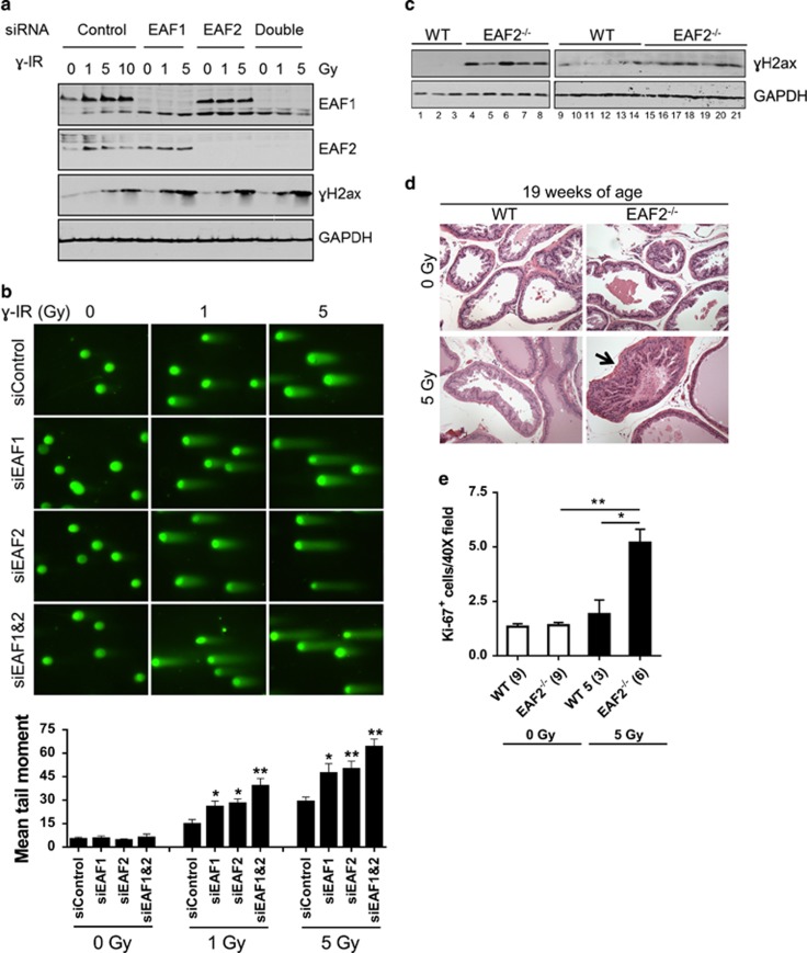
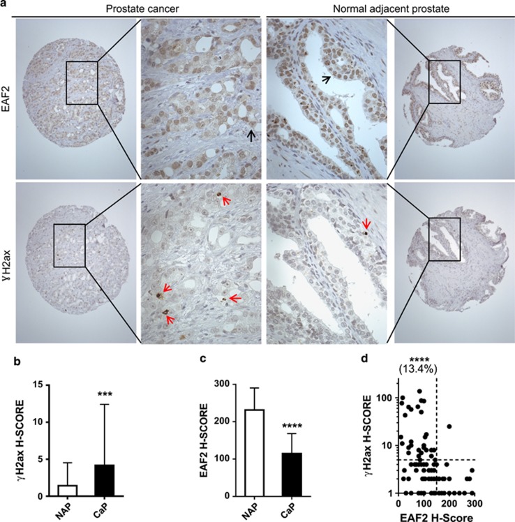
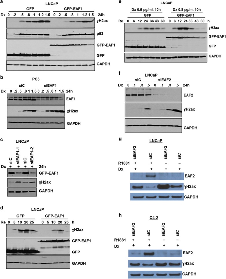
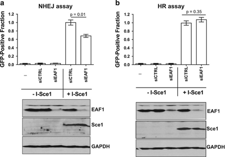
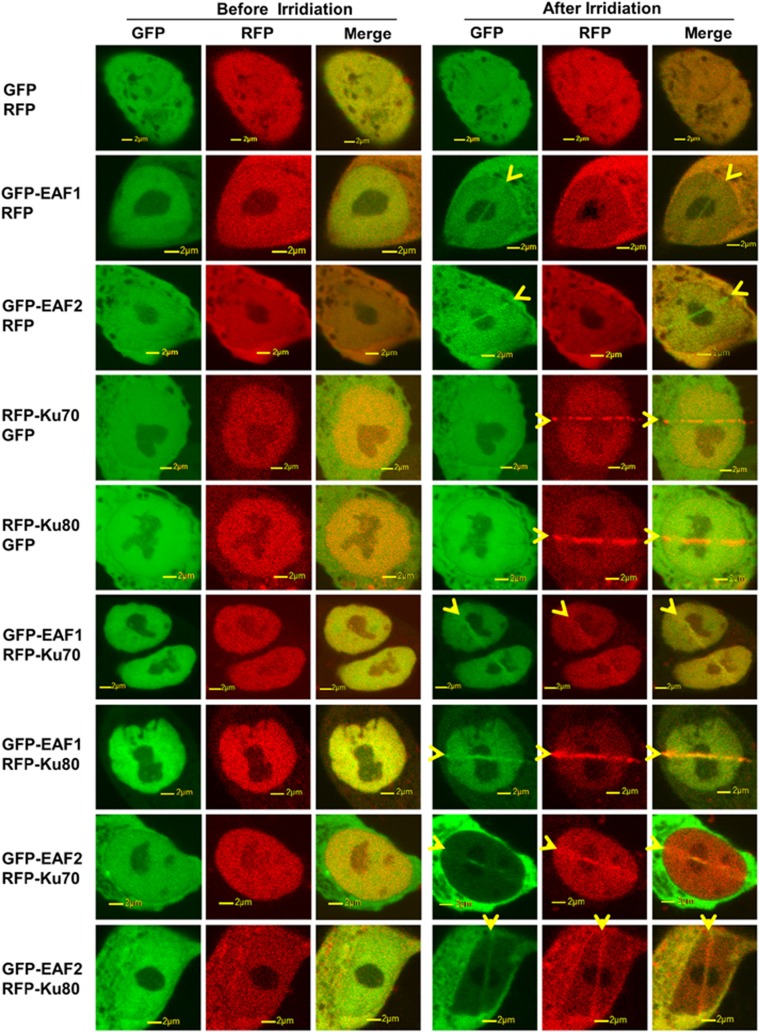
Androgens are known to protect prostate cancer cells from DNA damage. Recent studies showed regulation of DNA repair genes by androgen receptor signaling in prostate cancers. ELL-associated factor 2 (EAF2) is an androgen-regulated tumor suppressor and its intracellular localization can be modulated by ultraviolet light, suggesting a potential role for EAF2 in androgen regulation of DNA repair in prostate cancer cells. Here we show that knockdown of EAF2 or its homolog EAF1 sensitized prostate cancer cells to DNA damage and the sensitization did not require p53. EAF2 knockout mouse prostate was also sensitized to γ-irradiation. Furthermore, EAF2 knockdown blocked androgen repression of LNCaP or C4-2 cells from doxorubicin induction of γH2ax, a DNA damage marker. In human prostate cancer specimens, EAF2 expression was inversely correlated with the level of γH2ax. Further analysis showed that EAF2 and EAF1 are required for the recruitment and retention of Ku70/Ku80 to DNA damage sites and play a functional role in nonhomologous end-joining DNA repair. These findings provide evidence for EAF2 as a key factor mediating androgen protection of DNA damage via Ku70/Ku80 in prostate cancer cells.


Conflict of interest statement

The authors declare no conflict of interest.

Figures

Figure 1
Figure 1
Knockdown of EAF1 and EAF2 sensitizes prostatic cells to DNA damage. (a) Effect of siEAF1 and/or siEAF2 on γH2ax induction by γ-irradiation in LNCaP cells. Cells were irradiated at indicated dosages 48 h after siRNA knockdown and collected 5 h after irradiation. (b) Influence of siRNA knockdown of EAF1 and/or EAF2 on single-cell neutral gel electrophoresis (COMET) assay of LNCaP cells treated with γ-irradiation as in (a). Mean value of tail moments of cells with or without irradiation shown below. (c) Knockout of EAF2 sensitized prostate to γ-radiation-induced DNA damage. γH2ax expression in anterior prostate lobes of wild-type (WT) and EAF2−/− male mice at the age of 5–7 months at 24 h after γ-irradiation (8 Gy). Blots were reprobed with GAPDH (glyceraldehyde-3-phosphate dehydrogenase) antibody to provide loading control. Numbers indicate lanes. (d) Decreased latency of prostatic intraepithelial neoplasia (PIN) lesions (black arrow) induced by γ-radiation in EAF2−/− mice. (e) Ki-67 immunostaining of EAF2−/− mouse prostate tissues at 19 weeks of age treated with 5 Gy of γ-radiation at 8 weeks of age. *P<0.05, **P<0.01. See also Supplementary Figure S1.
Figure 2
Figure 2
Inverse correlation of EAF2 and γH2ax expression in human prostate cancer specimens. (a) Immunostaining of EAF2 and γH2ax in normal adjacent prostate and prostate cancer. Black and red arrows indicate staining of EAF2 and γH2ax, respectively. (b) Quantification of EAF2 immunostaining intensity H-Score in normal adjacent prostate (NAP) and prostate cancer (CaP) tissue specimens. (c) Quantification of γH2ax staining intensity H-Score in matched NAP and CaP tissue specimens (n=233). Scale bars: 100 μm, × 40. (d) Scatter plot of EAF2 and γH2ax immunostaining intensity in human prostate tissue specimens. Positive γH2ax staining (H-Score ⩾5) in specimens with EAF2 downregulation (H-Score <150) was significantly higher than in those with EAF2 expression (H-Score ⩾150) according to Fisher's exact test (P<0.0001). ***P<0.001, ****P<0.0001.
Figure 3
Figure 3
EAF1 enhances DNA repair and EAF2 knockdown inhibits androgen protection against doxorubicin-induced DNA damage. (a) LNCaP/GFP and LNCaP/GFP-EAF1 cells treated with indicated doses of doxorubicin (Dx, μg/ml) for 24 h. (b) Knockdown of EAF1 sensitized p53-negative PC3 cells to Dx-induced DNA damage. (c) After siRNA knockdown of EAF1 in LNCaP/GFP-EAF1 cells for 48 h, cells were treated with Dx (0.4 μg/ml) for additional 24 h. (d) LNCaP/GFP and LNCaP/GFP-EAF1 cells were treated with 0.4 μg/ml Dx for 12 h. Cells were then washed with PBS twice and cultured in fresh medium to recover (Re) for indicated time in hours. (e) LNCaP/GFP and LNCaP/GFP-EAF1 cells were treated with Dx (0.5 or 0.8 μg/ml) for 10 h and then allowed to recover as in (d). (f) LNCaP cells treated with siRNA targeting EAF2 for 48 h and then with Dx at indicated concentrations for an additional 24 h in the presence of 1 nM dihydrotestosterone (DHT) to enhance EAF2 expression. (g) LNCaP cells treated with siRNA targeting EAF2 in both the presence and absence of androgens (1 nM R1881). (h) C4-2 cells treated with siRNA targeting EAF2 in both the presence and absence of androgens (1 nM R1881). Cells were cultured in charcoal-stripped RPMI-1640 with and without 1 nM R1881 (g and h). LNCaP or C4-2 cells treated with siRNA targeting EAF2 for 48 h and then treated with 0.5 μg/ml of Dx for an additional 24 h. Blots were reprobed with GAPDH (glyceraldehyde-3-phosphate dehydrogenase) antibody to confirm equal protein loading.
Figure 4
Figure 4
Knockdown of EAF1 impairs NHEJ but not HR of double-strand breaks (DSBs) in chromosomal DNA. (a) Assay for NHEJ of chromosomal DSBs in H1299 (dA3-1) cells. Transiently expressed I-SceI protein cleaves the I-SceI sites and produces DSBs with incompatible ends in the substrate. NHEJ of two broken DNA ends of chromosomal DNA results in EGFP expression. GFP-positive fraction of cells treated with siEAF1 or siCTRL indicates frequency of NHEJ repair of chromosomal DNA. Western blot analysis was performed to confirm knockdown of EAF1 and Sce1 expression. (b) Assay for HR frequency of chromosomal DNA containing a recombination substrate DR-GFP in HeLa cells. GFP-positive fraction of cells treated with siEAF1 or siCTRL indicate frequency of HR repair of chromosomal DNA. Western blot confirmed knockdown of EAF1 and Sce1 expression. Blots were reprobed with GAPDH (glyceraldehyde-3-phosphate dehydrogenase) antibody to provide protein loading control.
Figure 5
Figure 5
Response and co-accumulation of EAF and Ku family proteins at laser microirradiation-induced DSBs sites. LNCaP cells were transiently transfected with GFP or GFP-tagged EAF1, EAF2 and red fluorescent protein (RFP) or RFP-tagged Ku70 and Ku80 expression vectors and treated with laser microirradiation to induce DSBs 24 h after transfection before and after irradiation. Accumulation of the transfected proteins was indicated by GFP (green) or RFP (red) fluorescence at laser-irradiated sites. Yellow arrowheads indicate direction of laser irradiation. Co-accumulation was visualized in merged images.
Figure 6
Figure 6
EAF1 and EAF2 bind to DNA repair proteins Ku70 and Ku80. (a) Co-immunoprecipitation of transfected EAF1 with Ku70 and Ku80. HEK293 cells were transiently transfected with the mammalian expression vectors for Myc-EAF1 and/or GFP-Ku70 and GFP-Ku80. Whole-cell lysates (WCLs) were immunoprecipitated with immobilized anti-GFP protein G beads and immunoblotted with anti-Myc or anti-GFP. (b) Co-immunoprecipitation of transfected EAF2 with Ku70 and Ku80 in HEK293 cells transiently transfected with mammalian expression vectors HA-EAF2 and/or GFP-Ku70 and GFP-Ku80. Cell lysates were immunoprecipitated with immobilized anti-GFP protein G beads and immunoblotted with anti-HA or anti-GFP. (c) Co-immunoprecipitation of endogenous EAF1 with Ku70 and Ku80. LNCaP cells were cultured in the presence of 1 nM dihydrotestosterone (DHT) for 24 h and then treated with and without doxorubicin (Dx, 1.0 μg/ml, 5 h) before harvest and nuclear extract preparation. Nuclear extracts (NEs) were precipitated using immobilized anti-EAF1 antibody and immunoblotted with anti-Ku70, anti-Ku80 and anti-EAF1 antibodies. (d) Co-immunoprecipitation of endogenous EAF2 with Ku70 and Ku80. The same LNCaP cell nuclear extracts as in (c) were precipitated using immobilized anti-EAF2 antibody and immunoblotted with anti-Ku70, anti-Ku80 and anti-EAF2 antibodies.
Figure 7
Figure 7
Accumulation of Ku70 and Ku80 proteins at laser microirradiation-induced damage sites after knockdown of EAF1 and/or EAF2. (a) Foci intensity of GFP-Ku70 accumulation at sites of laser microirradiation in LNCaP cells treated with indicated siRNA(s) for 48 h followed by transfection with GFP-Ku70 in the presence of 1 nM dihydrotestosterone (DHT) for 24 h. Mean foci intensity was measured every 20 s for 10 min and subtracted from background intensity. Right panel shows EAF1 and/or EAF2 knockdown and GFP-Ku70 expression. (b) Kinetics of GFP-Ku80 accumulation at laser-irradiated sites in LNCaP cells treated as in (a). Right panel shows EAF1 and/or EAF2 knockdown and GFP-Ku80 expression. (c) LNCaP cells were treated with siEAF1 for 48 h. Cells were then transfected with GFP-Ku70 alone or in combination of siEAF1-resistant GFP-EAF1 plasmids in the presence of 1 nM DHT. Kinetics of GFP-Ku70 accumulation at laser-irradiated sites was measured 24 h later. (d) Kinetics of GFP-Ku80 accumulation at laser-irradiated sites in LNCaP cells treated similarly to (d). (e) Protein expression of cells treated identically as in (c) and (d). Blots were reprobed with GAPDH (glyceraldehyde-3-phosphate dehydrogenase) antibody to confirm equal protein loading. See also Supplementary Figure S2.

References

    1. Siegel R, Naishadham D, Jemal A. Cancer statistics, 2013. CA Cancer J Clin 2013; 63: 11–30. - PubMed
    1. Bolla M, Van Tienhoven G, Warde P, Dubois JB, Mirimanoff RO, Storme G et al. External irradiation with or without long-term androgen suppression for prostate cancer with high metastatic risk: 10-year results of an EORTC randomised study. Lancet Oncol 2010; 11: 1066–1073. - PubMed
    1. Koontz BF, Lee WR. External beam radiation therapy for clinically localized prostate cancer: when and how we optimize with concurrent hormonal deprivation. Arch Esp Urol 2011; 64: 858–864. - PubMed
    1. Goodwin JF, Schiewer MJ, Dean JL, Schrecengost RS, de Leeuw R, Han S et al. A hormone-DNA repair circuit governs the response to genotoxic insult. Cancer Discov 2013; 3: 1254–1271. - PMC - PubMed
    1. Polkinghorn WR, Parker JS, Lee MX, Kass EM, Spratt DE, Iaquinta PJ et al. Androgen receptor signaling regulates DNA repair in prostate cancers. Cancer Discov 2013; 3: 1245–1253. - PMC - PubMed

Publication types

MeSH terms