. 2017 Apr;36(15):2160-2171.
doi: 10.1038/onc.2016.375. Epub 2016 Oct 10.

Evolutionarily conserved dual lysine motif determines the non-chaperone function of secreted Hsp90alpha in tumour progression

Affiliations

Evolutionarily conserved dual lysine motif determines the non-chaperone function of secreted Hsp90alpha in tumour progression

M Zou et al. Oncogene. 2017 Apr.

Erratum in

Abstract

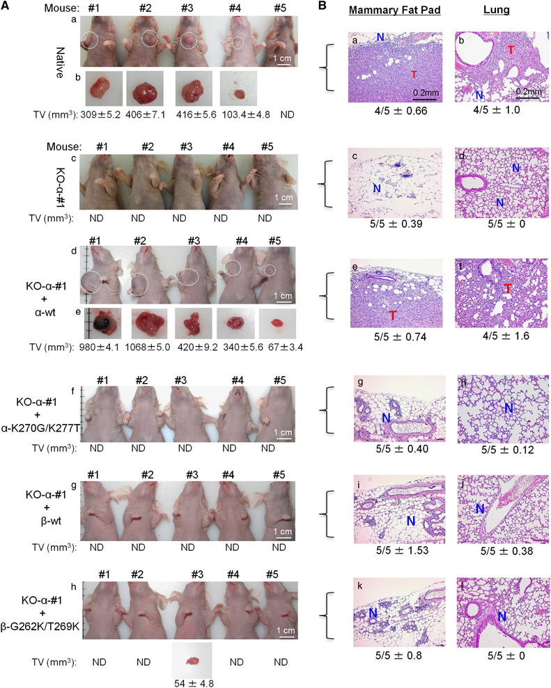
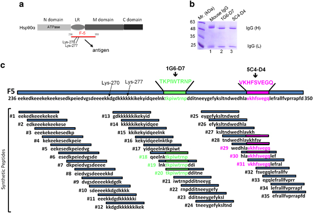
Both intracellular and extracellular heat shock protein-90 (Hsp90) family proteins (α and β) have been shown to support tumour progression. The tumour-supporting activity of the intracellular Hsp90 is attributed to their N-terminal ATPase-driven chaperone function. What molecular entity determines the extracellular function of secreted Hsp90 and the distinction between Hsp90α and Hsp90β was unclear. Here we demonstrate that CRISPR/Case9 knocking out Hsp90α nullifies tumour cells' ability to migrate, invade and metastasize without affecting the cell survival and growth. Knocking out Hsp90β leads to tumour cell death. Extracellular supplementation with recombinant Hsp90α, but not Hsp90β, protein recovers tumourigenicity of the Hsp90α-knockout cells. Sequential mutagenesis identifies two evolutionarily conserved lysine residues, lys-270 and lys-277, in the Hsp90α subfamily that determine the extracellular Hsp90α function. Hsp90β subfamily lacks the dual lysine motif and the extracellular function. Substitutions of gly-262 and thr-269 in Hsp90β with lysines convert Hsp90β to a Hsp90α-like protein. Newly constructed monoclonal antibody, 1G6-D7, against the dual lysine region of secreted Hsp90α inhibits both de novo tumour formation and expansion of already formed tumours in mice. This study suggests an alternative therapeutic approach to target Hsp90 in cancer, that is, the tumour-secreted Hsp90α, instead of the intracellular Hsp90α and Hsp90β.


Conflict of interest statement

The authors declare no conflict of interest.

Figures

Figure 1
Figure 1
Distinct roles for Hsp90α and Hsp90β in cancer cell survival and tumourigenicity. (A) Survival of MDA-MB-231 cells under drug selection following CRISPR-Cas9 Hsp90α gene (panels a, b and c) or Hsp90β gene (panels d, e and f) knockout was measured by counting the live cells over time (n=4). (B) Two Hsp90α-knockout clones (KO-α#1 and KO-α#2) showed complete absence of Hsp90α protein (panel g, lanes 3 and 4), in comparison with either short hairpin RNA (shRNA)-Hsp90α knockdown (lane 2) or the native MDA-MB-231 (lane 1) cells. (C) Specificity of the anti-Hsp90α and Hsp90β antibodies was confirmed by using the recombinant proteins in the Western. (D) Hsp90α gene knockout blocks Hsp90α (panel l), but not Hsp90β (panel m), secretion. (E) Growth curves of the native, shRNA-Hsp90α-knockdown (shRNA-α) and Hsp90α-knockout (KO-α-#1) MDA-MB-231 cells in the absence or presence of 10% foetal bovine serum (FBS). (F) Effect of Hsp90α gene on signalling in response to epidermal growth factor or TGFα.
Figure 2
Figure 2
Hsp90α knockout eliminates motility and invasiveness of the tumour cells. Serum-starved cells were subjected to colloidal gold motility (5000 cells per well) and the Matrigel invasion (20 000 cells per well) assays. (A) The migration tracks of MDA-MB-231, HBL-100 and human keratinocytes without (−) or with (+) 10% foetal bovine serum were quantitated by a computer-assisted analysis as MI (Materials and methods), as shown. (B) The motility measurements of the native MDA-MB-231, KO-α-#1 and KO-α-#2 cells in the absence (−) or presence (+) of human recombinant Hsp90α or human recombinant Hsp90β protein stimulation. (C) Matrigel invasion assay of the indicated cell lines with or without added human recombinant Hsp90α or human recombinant Hsp90β protein and the results quantitated as percentage of the invaded cells over the total number of seeded cells (Inv. %). (D) Evidence for FPLC-purified human recombinant Hsp90α and human recombinant Hsp90β proteins used in the above rescue experiments. (E) Evidence for downregulation of LRP-1 receptor in MDA-MB-231 cells by a lentivirus-delivered short hairpin RNA. (F) Matrigel invasion assay of LRP-1-downregulated cells in the absence or presence of human recombinant Hsp90α protein. Data were representative of multiple (n⩾ 4) independent experiments and represented as mean±s.e.m. P<0.05.
Figure 3
Figure 3
Lysine-270 and lysine-277 determine extracellular function of Hsp90α mutagenesis to identify the essential amino-acid residues for extracellular pro-motility activity of human Hsp90α. (a) Deletion mutagenesis narrowed the pro-motility activity of the 115-amino-acid F-5 fragment down to a 27-amino-acid peptide, F-8 fragment. (b) Comparison of F-8 from Hsp90α (F-8α) with the corresponding sequence of Hsp90β, F-8β, shows eight amino acids in F-8α (green) substituted with variant amino acids in F-8β (red). (c) Eight synthetic peptides with each of the eight amino acids in F-8α replaced with each of the corresponding amino-acid residues from F-8β (in red) were screened for their ability to rescue the motility defect of Hsp90α-knockout MDA-MB-231 cells, with F-8α and F-8β as positive and negative controls respectively. Quantitation of the cell motility is shown as MI (%). (d) K-270 and K-277 and their corresponding amino acids, G-262 and T-269, in Hsp90β were emphasized as focus of further studies.
Figure 4
Figure 4
Lysine-270 and lysine-277 substitutions convert Hsp90β to Hsp90α-like protein to promote cancer cell motility and invasion. (A) A schematic representation of lysine-270 and lysine-277 in full-length Hsp90α and creations of the Hsp90α-K270G/K277T and Hsp90α -D271K mutants. (B) A schematic representation of glycine-262 and threonine-269 in full-length Hsp90β and creations of the Hsp90β-G262K/T269K and Hsp90β-K263D mutants. (C) Sodium dodecyl sulfate–polyacrylamide gel electrophoresis (SDS–PAGE) with Coomassie Brilliant Blue staining of FPLC-purified recombinant Hsp90α and Hsp90β proteins. Commercial bovine serum albumin (BSA) is included as a comparison for relative quantities. (D) Circular dichroism (CD) revealed the secondary structural profiles of Hsp90α, Hsp90β, and their mutant proteins. (E) Rescue of the invasion defect of the KO-α-#1 cells (panel b vs panel a) by supplementation with wild-type Hsp90α (panel c), Hsp90α-G/T mutant (panel d), Hsp90α-D271K mutant (panel e), wild-type Hsp90β (panel f), Hsp90βK/K mutant (panel g) and Hsp90β-K263D mutant (panel h) (30 μg/ml). Quantitation of invasion (Inv. %) is included.
Figure 5
Figure 5
Tumour formation and metastasis in mice requires Lysine-270 and lysine-277 of Hsp90α. (A) Results of a representative experiment showing tumour formation of injected (5 × 10⁶) native MDA-MB-231 (panels a and b), KO-α-#1 cells alone (panel c) or KO-α-#1 cells supplemented with wild-type human recombinant Hsp90α (panels d and e), or with human recombinant Hsp90α-G/T mutant (panel f) or wild-type human recombinant Hsp90β (panel g) or with human recombinant Hsp90β-K/K mutant (panel h) protein to the mammary fat pad of nude mice (n=5 per group). The detected tumour formation was indicated by dotted circles and excised tumours measured for their Tumour Volume (TV, mm3). (B) Histological (H&E) analyses of the tumour, mammary fat pad tissue and the lung cryosections from non-tumour and tumour-containing mice as indicated. This experiment was repeated three times (n=4 or 5 per group). hr, human recombinant; T (red), tumour; N (blue), normal tissue; 5/5, five out of the five mice; Data are represented as mean±s.e.m. P<0.05.
Figure 6
Figure 6
Development of secreted Hsp90α-neutralizing monoclonal antibodies against the dual lysine region of human Hsp90α. (a) A schematic illustration of the antigen (F-5) location in reference to the dual lysine residues in full-length Hsp90α. (b) IgG was purified by protein-G Sepharose affinity chromatography from two hybridoma clones, 1G6-D7 and 5C4-D4, and analysed by sodium dodecyl sulfate–polyacrylamide gel electrophoresis (SDS–PAGE) with coomassie brilliant blue staining. Regular mouse IgG was used as a control. (c) 1G6-D7 (IgG1κ) and 5C4-D4 (IgG2aκ) are mapped to bind to TKPIWTRNP and VKHFSVEGQ, respectively, within the antigen.
Figure 7
Figure 7
1G6-D7 blocks tumour formation and metastasis in vivo. Although 1G6-D7 inhibits tumour cell invasion in vitro (see Supplementary Figure S6), it also works in vivo. (A) A representative experiment showing tumour formation of injected native MDA-MB-231 cells (5 × 10⁶) in mice (n=3 or 5 per group) that were intravenously administered with either control mouse IgG or 1G6-D7 (5 mg/kg/injection) (Methods). (B) Measurement of tumour volumes on live mice over 4 weeks. (C) H&E staining of the primary tumour/tissue (panels a and c) or the lungs (panels b and d) sections from IgG control- or 1G6-D7-treated mice as indicated. T, tumour; N, normal tissue. 5/5, five out of five mice per group. This experiment was repeated three times (n=4 or 5 mice per group). Data are represented as mean±s.e.m. P<0.05.
Figure 8
Figure 8
1G6-D7 inhibits expansion of pre-formed tumours in vivo. (A) A representative experiment showing tumour formation by injected parental MDA-MB-231 cells (5 × 10⁶) over 35 days. On day 5 with an average tumour size of 60 mm3, either vehicle, control mouse IgG or 1G6-D7 was injected via IV (5 mg/kg) and around the tumour site (125 μg per injection). Measurement of the tumour volumes on live mice (n=4 or 5) was carried out every 5 days. This experiment was repeated twice. Data are represented as mean±s.e.m. P<0.05. (B) The images of the mice and the TV measurement of the excised tumours from the mice on day 36 (the s.d. represents three independent measurements with different angles of a tumour). (C) Based on the previously evaluated crystal structures of Hsp90α’s NTD (green) and MD (blue) plus CTD (red) domains and their fit into a cryoEM map of full-length Hsp90α bound to Hop (MODELLERv9.14). Lysine-270 and lysine-277 are located in the unstructured linker region (LR) between the NTD and MD domains, called F-5. Inhibitors, such as 1G6-D7, targeting the dual lysine residues (larger box) could block secreted Hsp90α-triggered tumourigenesis.

References

    1. Young JC, Moarefi I, Hartl FU . Hsp90: a specialized but essential protein-folding tool. J Cell Biol 2001; 154: 267–273. - PMC - PubMed
    1. Sahu D, Zhao Z, Tsen F, Cheng CF, Park R, Situ AJ et al. A potentially common peptide target in secreted heat shock protein-90α for hypoxia-inducible factor-1α-positive tumours. Mol Biol Cell 2012; 23: 602–613. - PMC - PubMed
    1. Dong H, Zou M, Bhatia A, Jayaprakash P, Hofman F, Yin Q, Chen M, Woodley DT, Li W . Breast cancer MDA-MB-231 cells use secreted heat shock protein-90alpha (Hsp90α) to survive a hostile hypoxic environment. Sci Rep 2016; 6: 20605. - PMC - PubMed
    1. Obermann WM, Sondermann H, Russo AA, Pavletich NP, Hartl FU . In vivo function of Hsp90 is dependent on ATP binding and ATP hydrolysis. J Cell Biol 1998; 143: 901–910. - PMC - PubMed
    1. Drysdale MJ, Brough PA, Massey A, Jensen MR, Schoepfer J . Targeting Hsp90 for the treatment of cancer. Curr Opin Drug Discov Devel 2006; 9: 483–495. - PubMed