Review
. 2016 Dec;14(12):744-759.
doi: 10.1038/nrmicro.2016.141. Epub 2016 Oct 10.

Treponema pallidum, the syphilis spirochete: making a living as a stealth pathogen

Affiliations
Review

Treponema pallidum, the syphilis spirochete: making a living as a stealth pathogen

Justin D Radolf et al. Nat Rev Microbiol. 2016 Dec.

Abstract

The past two decades have seen a worldwide resurgence in infections caused by Treponema pallidum subsp. pallidum, the syphilis spirochete. The well-recognized capacity of the syphilis spirochete for early dissemination and immune evasion has earned it the designation 'the stealth pathogen'. Despite the many hurdles to studying syphilis pathogenesis, most notably the inability to culture and to genetically manipulate T. pallidum, in recent years, considerable progress has been made in elucidating the structural, physiological, and regulatory facets of T. pallidum pathogenicity. In this Review, we integrate this eclectic body of information to garner fresh insights into the highly successful parasitic lifestyles of the syphilis spirochete and related pathogenic treponemes.


Conflict of interest statement

The authors report no conflicts of interest.

Figures
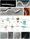
Figure 1
Figure 1. Morphology and cell envelope architecture of T. pallidum, the stealth pathogen
A. Darkfield micrograph showing the flat wave morphology of T. pallidum. Asterisks and arrowhead indicate segments oriented 90 degrees from each other (with permission from Reference 15). B. Top view of a surface-rendered model of T. pallidum generated from cryoelectron tomograms showing the outer and cytoplasmic membranes (transparent yellow), flagellar motors (basal bodies, dark lavender), flagellar filaments (light lavender), cytoplasmic filaments (orange), cap (green), and cone (pink) (with permission from Reference 15). C. Freeze-fracture electron microscopy reveals scarce particles (integral membrane proteins) within the T. pallidum outer membrane. Convex and concave leaflets of the outer membrane are indicated; arrowheads indicate particles in the two leaflets (with permission from Reference 155). D. Scanning probe microscopy reveals rare particles on the T. pallidum surface (arrows), often located on the bulge in the outer membrane created by the underlying periplasmic flagella (asterisks) (with permission from Reference 91). E. Model for the molecular architecture of the T. pallidum cell envelope. Shown in the outer membrane are BamA (TP0326),, a generic bipartite (that is, full-length) Tpr attached by its N-terminal portion to the peptidoglycan , a generic non-Tpr β-barrel, and TP0453 (p30.5), a structurally characterized lipoprotein attached to the outer membrane inner leaflet. Tprs, such as TprF, lacking the MOSPC β-barrel forming domain, are located in the periplasm. Substrates present in high concentration in the extracellular milieu probably traverse the outer membrane by simple diffusion through porins, such as TprC. Prototypic ABC-like transporters use a periplasmic substrate-binding protein (SBP), typically lipoproteins, and components with transmembrane and ATP-binding domains. The energy coupling factor (ECF)-type ABC transporters uses a transmembrane ligand-binding protein in place of a separate periplasmic SBP. To substitute for ATP hydrolysis, the symporters use a transmembrane permease that relies on energy from a chemiosmotic or electrochemical gradient that may be generated by the Rnf complex. The tripartite ATP-independent periplasmic (TRAP) transporters also lack ATP-binding modules and use transmembrane electrochemical gradients, but they are more complex. T. pallidum seems to have evolved a variation on the TRAP theme by also using an additional periplasmic component protein (TatT (TP0956)) containing a tetratricopeptide repeat (TPR) motif, giving rise to a newly described system denoted as a TPR protein-associated TRAP transporter (TPAT). The TPR protein TatT likely associates with the SBP TatP (TP0957) in a heterohexameric fashion to carry out ligand binding and uptake; structural analyses suggest that this complex may accommodate a chain-like hydrophobic molecule(s), such as a long-chain fatty acid(s). Uptake likely is facilitated by a putative membrane permease (TatQ-M (TP0958)) of the TPAT system,. F. Cryotomographic section of T. pallidum near the cell end showing the peptidoglycan layer (arrowheads) midway between the outer membrane and cytoplasmic membrane (with permission from Reference 15). G. Cryotomographic section of T. pallidum showing chemoreceptor arrays (arrows, with permission from Reference 91). H. Cryotomographic slice showing the cone-shaped structure at T. pallidum cell ends (with permission from Reference 15) along with darkfield micrograph of T. pallidum stably attached via its tip to the surface of a trophoblast cell. White arrowheads indicate fine fibers between the cone and the outer membrane.
Figure 2
Figure 2. Energy generation, amino acid biosynthesis, and regeneration of NAD+ in T. pallidum
T. pallidum’s glycolytic and pyruvate-to-acetate fermentation pathways are coupled to a putative Rnf pump, resulting in production of ATP by an ATP synthase; phosphoenolpyruvate (PEP) from glycolysis can be further metabolized to amino acids via PEP carboxykinase (PEP-CK). Metabolites, enzymes, and cofactors are shown in green, red, and blue, respectively. Abbreviations for enzymes are as follows: PPFK, phosphofructokinase; Nox, NADH oxidase; PPDK, pyruvate phosphate dikinase; OAD, oxaloacetate decarboxylase; TpaaT, aspartate aminotransferase; AsnA, asparagine synthetase; LDH, D-lactate dehydrogenase; PFOR, pyruvate-flavodoxin oxidoreductase; Pta, phosphate acetyl transferase; AckA, acetate kinase. Abbreviations for compounds/metabolites are as follows: Fru-6-P, fructose 6-phosphate; Fru-1, 6-P2, fructose 1, 6-bisphosphate; Pi, inorganic phosphate; PPi, inorganic pyrophosphate; CoA, coenzyme A; Acetyl-P; acetyl phosphate; RNF, a putative chemiosmotic pump with similarities to the Rhodobacter nitrogen fixation complex; Flavodoxinox and Flavodoxinred, oxidized and reduced forms of flavodoxin.
Figure 3
Figure 3. Proposed pathways for control of alternative sigma factors in T. pallidum
A. σE cell envelope stress responses in T. pallidum and E. coli. Binding of misfolded OMPs to the PDZ domain of DegS (TP0773) initiates transduction of the stress signal across the cytoplasmic membrane. T. pallidum lacks a recognizable ortholog for RseB, a negative regulator that binds to RseA and protects it from DegS-mediated cleavage. B. σ (RpoN)-dependent gene expression. T. pallidum contains two enhancer binding proteins, the NtrC-like TP0519 and the NorR-like TP0082. Because TP0519 lacks a cognate histidine kinase, phosphorylation of TP0519 may occur through an acetyl-phosphate donor. Activation of TP0082 is predicted to occur following the binding of a small molecule to its GAF domain. tpF1 (tp1038), encoding a bacterioferritin, and tp1012, encoding an RpoS-like sigma factor, are predicted to be transcribed by σ.
Figure 4
Figure 4. Domain architecture of the T. pallidum repeat (Tpr) family of proteins Tprs in T. pertenue (TPE) and T. pallidum (TPA) strains
The Tprs are divided into three subfamilies based on sequence relatedness; within subfamilies I and II, members are closely related to each other, whereas members of subfamily III are heterogeneous,. Full-length Tprs consist of: (i) a variable extreme N-terminal stretch (~50 amino acids); (ii) MOSPN, a conserved N-terminal domain related to the corresponding domain in the N-terminus of the major outer sheath protein (MOSP) of T. denticola; (iii) a variable central region; and (iv) MOSPC, a conserved C-terminal β-forming domain related to the corresponding domain in T. denticola MOSP. The thin yellow lines denote amino acid variation in TPE paralogs relative to the corresponding paralogs in the TPA Nichols strain. Shown in umber are insertions in the MOSPC domains of TprG and TprJ relative to the corresponding paralog in the TPA Nichols reference strain. The central domain of TprJ in TPE contains sequences derived from the corresponding region of TprG. TprL in the TPA Nichols strain contains an extended variable N-terminal stretch. In TPE, TprA and TprF are full-length, bipartite proteins. The blue and red lines, respectively, are used to indicate sequence variation in TprD2 and TprI relative to TprC/D in TPA Nichols. With the exception of the Sea81-4 TPA strain, TprA is truncated, lacking the MOSPC domain; TprF is truncated in all TPA strains. TprK is not shown because of its hypervariability.

Similar articles

Cited by

References

    1. Giacani L, Lukehart SA. The endemic treponematoses. Clin Microbiol Rev. 2014;27:89–115. This is a contemporary review of the origins, global epidemiology, and clinical features of the novenereal treponematoses and their continuing threat to worldwide health. - PMC - PubMed
    1. Sena AC, Pillay A, Cox DL, Radolf JD. In: Manual of Clinical Microbiology. Jorgensen JH, et al., editors. ASM Press; Washington, D.C: 2015. pp. 1055–1081.
    1. Miao R, Fieldsteel AH. Genetics of Treponema: relationship between Treponema pallidum and five cultivable treponemes. J Bacteriol. 1978;133:101–7. - PMC - PubMed
    1. Miao RM, Fieldsteel AH. Genetic relationship between Treponema pallidum and Treponema pertenue, two noncultivable human pathogens. J Bacteriol. 1980;141:427–9. - PMC - PubMed
    1. Smajs D, Norris SJ, Weinstock GM. Genetic diversity in Treponema pallidum: implications for pathogenesis, evolution and molecular diagnostics of syphilis and yaws. Infect Genet Evol. 2012;12:191–202. This is a comprehensive review of the genetic and evolutionary relatioships among pathogenic treponemes based on genomic sequencing data. - PMC - PubMed

MeSH terms

Substances