

Human MutL homolog 1 immunoexpression in oral leukoplakia and oral squamous cell carcinoma: A prospective study in Indian population
- PMID: 27721611
- PMCID: PMC5051294
- DOI: 10.4103/0973-029X.190948
Human MutL homolog 1 immunoexpression in oral leukoplakia and oral squamous cell carcinoma: A prospective study in Indian population
Abstract
Background: Mammalian mismatch repair system is responsible for maintaining genomic stability during repeated duplications, and human MutL homolog 1 (hMLH1) protein constitutes an important part of it. Various isolated studies have reported the altered expression of hMLH1 in oral leukoplakia (OL) and oral squamous cell carcinoma (OSCC). Research is lacking in the quantitative estimation and comparison of hMLH1 expression in OL and OSCC.
Aims: To evaluate, quantify and compare hMLH1 immunoexpression in normal oral mucosa, OL and OSCC.
Settings and design: Thirty patients of OL and thirty patients of OSCC formed the study group and thirty patients were included in the control group (normal oral mucosa). Formalin-fixed paraffin wax blocks were prepared from the tissue samples.
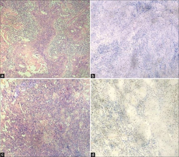
Materials and methods: Immunohistochemistry for hMLH1 was performed, and the total number of positive cells was counted in high-power fields, and based on that percentage positivity of hMLH1 was calculated in all the cases.
Statistical analysis: Kruskal-Wallis and t-test were used. P < 0.05 was considered to be statistically significant.
Results: The mean hMLH1 value in control group, leukoplakia and OSCC was 78.26, 54.33 and 40.97 respectively. hMLH1 immunoexpression showed decreasing indexes from control group to leukoplakia and then further to OSCC. hMLH1 expression was significantly lower in OSCC as compared to leukoplakia. There was no significant correlation of mean hMLH1 expression between different clinical and histopathological stages of leukoplakia and OSCC.
Conclusions: hMLH1 immunoexpression was inversely related to the degree of dysplasia. These findings suggest that there is a progressive decrease in hMLH1 expression from control to leukoplakia and further to OSCC. Thus, it can be concluded that hMLH1 can be used as a reliable biomarker for malignant transformation.
Keywords: Mismatch repair system; oral leukoplakia; oral squamous cell carcinoma.
Figures


References
-
- de Camargo Cancela M, Voti L, Guerra-Yi M, Chapuis F, Mazuir M, Curado MP. Oral cavity cancer in developed and in developing countries: Population-based incidence. Head Neck. 2010;32:357–67. - PubMed
-
- Warnakulasuriya S, Johnson NW, van der Waal I. Nomenclature and classification of potentially malignant disorders of the oral mucosa. J Oral Pathol Med. 2007;36:575–80. - PubMed
-
- van der Waal I. Potentially malignant disorders of the oral and oropharyngeal mucosa; present concepts of management. Oral Oncol. 2010;46:423–5. - PubMed
LinkOut - more resources
Full Text Sources
Other Literature Sources
