Review
. 2016 Sep 23:7:1503.
doi: 10.3389/fmicb.2016.01503. eCollection 2016.

Components of Adenovirus Genome Packaging

Affiliations
Review

Components of Adenovirus Genome Packaging

Yadvinder S Ahi et al. Front Microbiol. .

Abstract

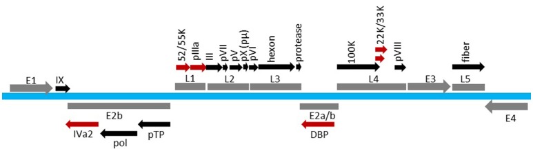
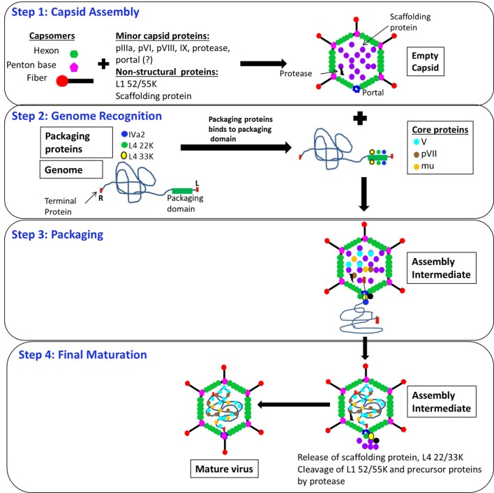
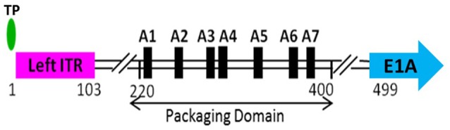
Adenoviruses (AdVs) are icosahedral viruses with double-stranded DNA (dsDNA) genomes. Genome packaging in AdV is thought to be similar to that seen in dsDNA containing icosahedral bacteriophages and herpesviruses. Specific recognition of the AdV genome is mediated by a packaging domain located close to the left end of the viral genome and is mediated by the viral packaging machinery. Our understanding of the role of various components of the viral packaging machinery in AdV genome packaging has greatly advanced in recent years. Characterization of empty capsids assembled in the absence of one or more components involved in packaging, identification of the unique vertex, and demonstration of the role of IVa2, the putative packaging ATPase, in genome packaging have provided compelling evidence that AdVs follow a sequential assembly pathway. This review provides a detailed discussion on the functions of the various viral and cellular factors involved in AdV genome packaging. We conclude by briefly discussing the roles of the empty capsids, assembly intermediates, scaffolding proteins, portal vertex and DNA encapsidating enzymes in AdV assembly and packaging.

Keywords: ATPase; IVa2; L4 22K; L4 33K; adenovirus; genome packaging; packaging domain; portal vertex.


Figures

Figure 1
Figure 1
Transcription map of HAdV-C5 genome. Viral genome is shown in blue. The early regions (E1, E2, E3, and E4) and the late regions (L1, L2, L3, L4, and L5) are shown in gray. Proteins expressed from various regions are indicated above or below the region. Factors involved in AdV packaging are shown in red. The map is not drawn to scale.
Figure 2
Figure 2
Predicted model of AdV assembly and packaging. Step 1: Capsid assembly: Empty capsids are assembled from hexon trimers and pentons, along with the minor capsid proteins (pIIIa, pVI, pVIII, IX, protease and portal) and the non-structural proteins (L1 52/55K and the scaffolding protein). Step 2: Genome recognition: Packaging proteins IVa2, L4 33K, L1 52/55K, and L4 22K bind to the packaging domain located close to the left end of the genome. The genome is associated with pTP at each end. Step 3: Packaging: The genome is inserted into the empty capsids through an unknown portal located at the unique vertex by the action of IVa2, L4 22K, and L4 33K. The core proteins V, VII and mu are incorporated into the empty capsids during or after genome incorporation. Step 4: Final maturation: Scaffolding protein (not known), L4 22K and 33K are released during or after genome incorporation. The virus encoded protease cleaves its substrates pIIIa, L1 52/55K, pVI, pVII, pVIII, mu and pTP, resulting in conformational changes in the capsid structure and maturation of the virus particle. pVI, precursor VI; pVII, precursor VII; pVIII, precursor VIII; pTP, precursor terminal protein.?, indicates that the identity is not known. L and R indicate left and right ends of the viral genome, respectively.
Figure 3
Figure 3
Schematic depiction of the left end of HAdV-C5 genome. The packaging domain of HAdV-C5 is located between 220 and 400 bp of the viral genome and consists of seven AT-rich repeats, referred to as A-repeats. The terminal protein (TP) is covalently attached to the 5′-phosphate. The left inverted terminal repeat (left ITR) extends from 1 to 103 bp. Transcription from the early region 1A (E1A) begins at 499 bp.
Figure 4
Figure 4
Known functional domains of IVa2 protein. Known functional domains of HAdV-C5 IVa2 protein (1–449 residues) are depicted. The locations of the nucleolar localization signal (NuLS), Walker A motif (A), Walker B motif (B), and nuclear localization signal (NLS) are shown. Dotted arrows (79–98, and 421–449) indicate the regions that are part of the bipartite DNA binding domain.

Similar articles

Cited by

References

    1. Ahi Y. S., Vemula S. V., Hassan A. O., Costakes G., Stauffacher C., Mittal S. K. (2015). Adenoviral L4 33K forms ring-like oligomers and stimulates ATPase activity of IVa2: implications in viral genome packaging. Front. Microbiol. 6:318. 10.3389/fmicb.2015.00318 - DOI - PMC - PubMed
    1. Ahi Y. S., Vemula S. V., Mittal S. K. (2013). Adenoviral E2 IVa2 protein interacts with L4 33K protein and E2 DNA-binding protein. J. Gen. Virol. 94(Pt 6), 1325–1334. 10.1099/vir.0.049346-0 - DOI - PubMed
    1. Ali H., LeRoy G., Bridge G., Flint S. J. (2007). The adenovirus L4 33-kilodalton protein binds to intragenic sequences of the major late promoter required for late phase-specific stimulation of transcription. J. Virol. 81, 1327–1338. 10.1128/jvi.01584-06 - DOI - PMC - PubMed
    1. Andrade F., Fellows E., Jenne D. E., Rosen A., Young C. S. (2007). Granzyme H destroys the function of critical adenoviral proteins required for viral DNA replication and granzyme B inhibition. EMBO J. 26, 2148–2157. 10.1038/sj.emboj.7601650 - DOI - PMC - PubMed
    1. Ariga H., Klein H., Levine A. J., Horwitz M. S. (1980). A cleavage product of the adenovirus DNA binding protein is active in DNA replication in vitro. Virology 101, 307–310. - PubMed