Review
. 2016:2016:4971572.
doi: 10.1155/2016/4971572. Epub 2016 Sep 19.

Applications of Anterior Segment Optical Coherence Tomography in Cornea and Ocular Surface Diseases

Affiliations
Review

Applications of Anterior Segment Optical Coherence Tomography in Cornea and Ocular Surface Diseases

Sang Beom Han et al. J Ophthalmol. 2016.

Abstract

Optical coherence tomography (OCT) is a noncontact technology that produces high-resolution cross-sectional images of ocular tissues. Anterior segment OCT (AS-OCT) enables the precise visualization of anterior segment structure; thus, it can be used in various corneal and ocular surface disorders. In this review, the authors will discuss the application of AS-OCT for diagnosis and management of various corneal and ocular surface disorders. Use of AS-OCT for anterior segment surgery and postoperative management will also be discussed. In addition, application of the device for research using human data and animal models will be introduced.


Conflict of interest statement

None of the authors have proprietary interests in the study or financial interests to disclose.
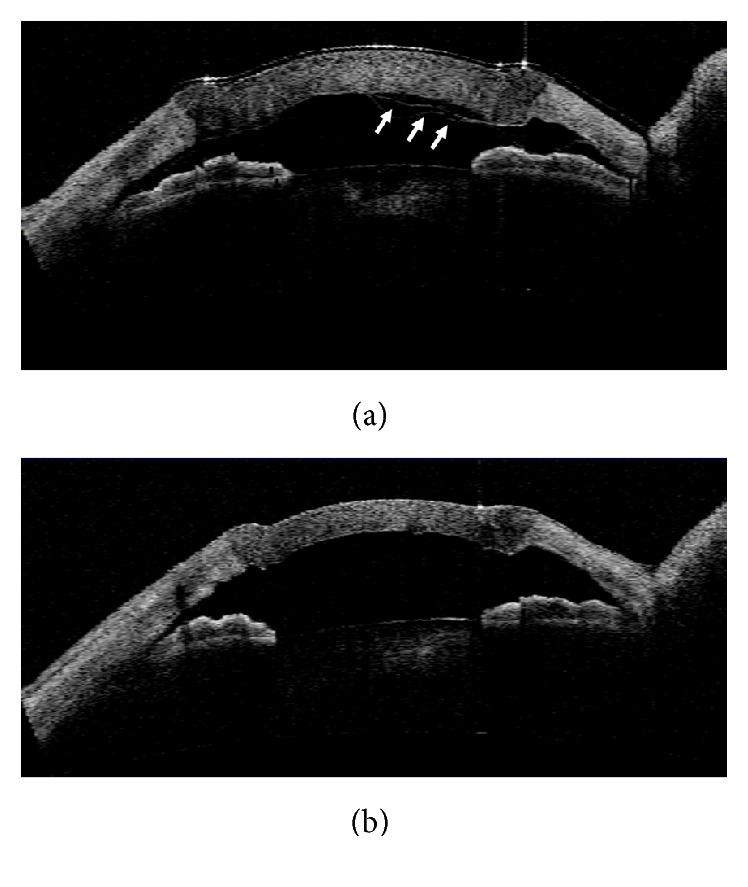
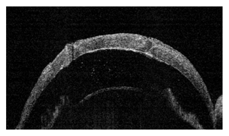
Figures

Figure 1
Figure 1
An AS-OCT image of a patient with granular dystrophy. AS-OCT can accurately show the location and depth of the visually significant stromal haze.
Figure 2
Figure 2
A TD AS-OCT image demonstrating localized Descemet's membrane detachment (white arrow) adjacent to the wound and internal wound gaping (black arrow) that was not detected in slit-lamp examination.
Figure 3
Figure 3
An AS-OCT image of precut donor cornea for Descemet's stripping automated endothelial keratoplasty (DSAEK) taken for evaluation of thickness of graft donor tissue.
Figure 4
Figure 4
An intraoperative AS-OCT image taken during deep anterior lamellar keratoplasty (DALK) that confirms an optimal descemetic or predescemetic dissection.
Figure 5
Figure 5
AS-OCT images demonstrating detachment of the Descemet's membrane (DM; white arrow) after DALK (a) and reattachment of the DM after intracameral air injection (b).
Figure 6
Figure 6
An AS-OCT image demonstrating anterior chamber inflammation in a rejected graft in a rat model.

References

    1. Huang D., Swanson E. A., Lin C. P., et al. Optical coherence tomography. Science. 1991;254(5035):1178–1181. doi: 10.1126/science.1957169. - DOI - PMC - PubMed
    1. Izatt J. A., Hee M. R., Swanson E. A., et al. Micrometer-scale resolution imaging of the anterior eye in vivo with optical coherence tomography. Archives of Ophthalmology. 1994;112(12):1584–1589. doi: 10.1001/archopht.1994.01090240090031. - DOI - PubMed
    1. Radhakrishnan S., Rollins A. M., Roth J. E., et al. Real-time optical coherence tomography of the anterior segment at 1310 nm. Archives of Ophthalmology. 2001;119(8):1179–1185. doi: 10.1001/archopht.119.8.1179. - DOI - PubMed
    1. Ramos J. L. B., Li Y., Huang D. Clinical and research applications of anterior segment optical coherence tomography—a review. Clinical and Experimental Ophthalmology. 2009;37(1):81–89. doi: 10.1111/j.1442-9071.2008.01823.x. - DOI - PMC - PubMed
    1. Wang J., Abou Shousha M., Perez V. L., et al. Ultra-high resolution optical coherence tomography for imaging the anterior segment of the eye. Ophthalmic Surgery, Lasers & Imaging. 2011;42(supplement):S15–S27. doi: 10.3928/15428877-20110627-02. - DOI - PubMed

LinkOut - more resources