

Dorsoscapularis triangularis: embryological and phylogenetic characterization of a rare variation of trapezius
- PMID: 27722016
- PMCID: PMC5052232
- DOI: 10.5115/acb.2016.49.3.213
Dorsoscapularis triangularis: embryological and phylogenetic characterization of a rare variation of trapezius
Abstract
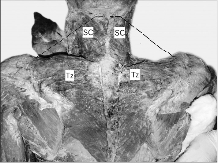
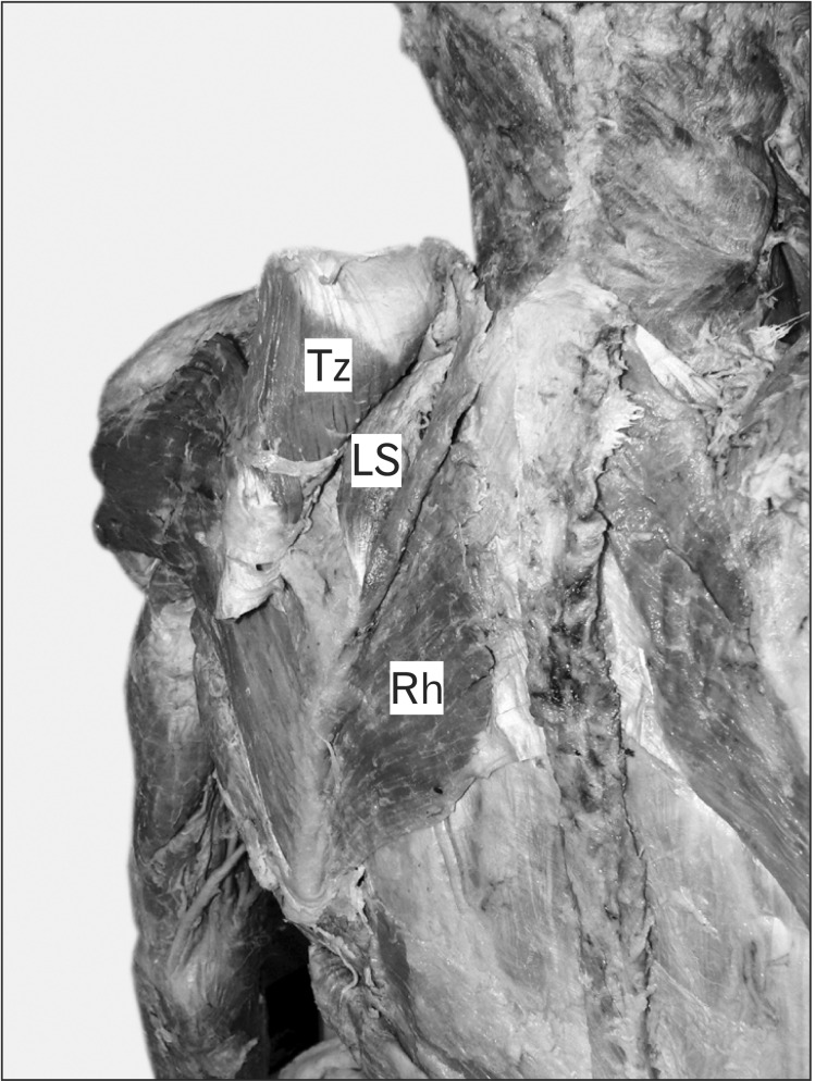
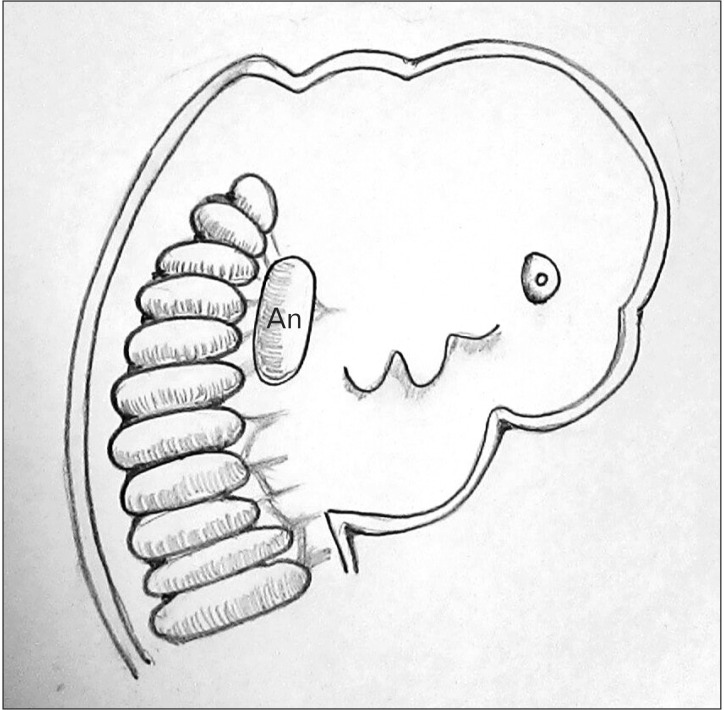
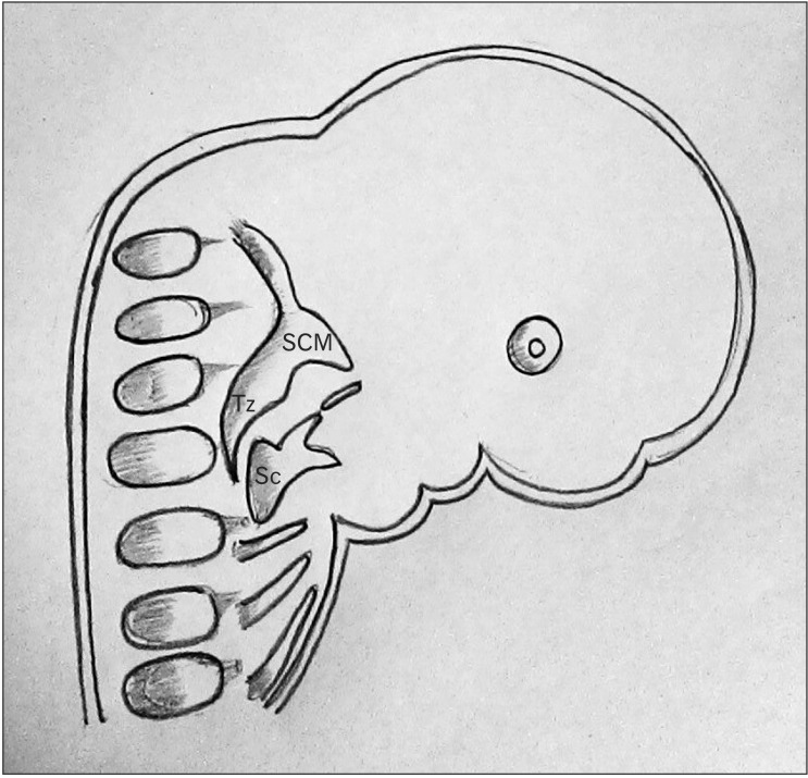
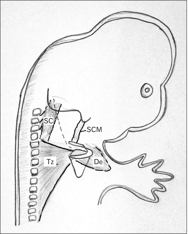
The muscle trapezius shows considerable morphological diversity. Variations include an anomalous origin and complete or partial absence of the muscle. The present study reported, a hitherto undocumented complete bilateral absence of the cervical part of trapezius. Based on its peculiar origin and insertion, it was named dorsoscapularis triangularis. The embryological, phylogenetic and molecular basis of the anomaly was elucidated. Failure of cranial migration of the trapezius component of the branchial musculature anlage to gain attachment on the occipital bone, cervical spinous processes, ligamentum nuchae between 11 mm and 16 mm stage of the embryo, resulted in this anomaly. A surgeon operating on the head and neck region or a radiologist analyzing a magnetic resonance imaging of the cervical region would find the knowledge of this morphological variation of trapezius useful in making clinical decisions.
Keywords: HOX D4; Molecular; Morphology; Trapezius; Variation.
Figures
Similar articles
-
Absence of Posterior Triangle: Clinical and Embryological Perspective.J Clin Diagn Res. 2017 Feb;11(2):AD01-AD02. doi: 10.7860/JCDR/2017/23896.9176. Epub 2017 Feb 1. J Clin Diagn Res. 2017. PMID: 28384846 Free PMC article.
-
Clinical anatomy of ligamentum nuchae.Clin Anat. 2003 Nov;16(6):484-93. doi: 10.1002/ca.10121. Clin Anat. 2003. PMID: 14566894
-
A macroscopical study of the trapezius muscle of sharks, with reference to the topographically related nerves and vein.Anat Anz. 1988;165(1):7-21. Anat Anz. 1988. PMID: 3358535
-
The spinal accessory nerve plexus, the trapezius muscle, and shoulder stabilization after radical neck cancer surgery.Ann Surg. 1988 Nov;208(5):654-61. doi: 10.1097/00000658-198811000-00019. Ann Surg. 1988. PMID: 3056289 Free PMC article. Review.
-
A case of left-sided absence and right-sided fenestration of the external jugular vein and a review of the literature.Surg Radiol Anat. 2015 Sep;37(7):883-6. doi: 10.1007/s00276-014-1398-z. Epub 2014 Nov 30. Surg Radiol Anat. 2015. PMID: 25432662 Review.
Cited by
-
Bilateral sternocleidomastoid variant with six distinct insertions along the superior nuchal line.Anat Cell Biol. 2018 Dec;51(4):305-308. doi: 10.5115/acb.2018.51.4.305. Epub 2018 Dec 29. Anat Cell Biol. 2018. PMID: 30637167 Free PMC article.
References
-
- Standring S. Gray's anatomy: the anatomical basis of clinical practice. 40th ed. Madrid: Elsevier Churchill Livingstone; 2008.
-
- Colletti G, Tewfik K, Bardazzi A, Allevi F, Chiapasco M, Mandalà M, Rabbiosi D. Regional flaps in head and neck reconstruction: a reappraisal. J Oral Maxillofac Surg. 2015;73:571.e1–571.e10. - PubMed
-
- Allouh M, Mohamed A, Mhanni A. Complete unilateral absence of trapezius muscle. McGill J Med. 2004;8:31–33.
-
- Horan FT, Bonafede RP. Bilateral absence of the trapezius and sternal head of the pectoralis major muscles: a case report. J Bone Joint Surg Am. 1977;59:133. - PubMed
-
- Yiyit N, Işıtmangil T, Öksüz S. Clinical analysis of 113 patients with Poland syndrome. Ann Thorac Surg. 2015;99:999–1004. - PubMed
Publication types
LinkOut - more resources
Full Text Sources
Other Literature Sources