. 2016 Dec;74(4):527-535.
doi: 10.1007/s12013-016-0758-1. Epub 2016 Oct 8.

The Effect of Substrate Stiffness on Cardiomyocyte Action Potentials

Affiliations

The Effect of Substrate Stiffness on Cardiomyocyte Action Potentials

Sean D Boothe et al. Cell Biochem Biophys. 2016 Dec.

Abstract

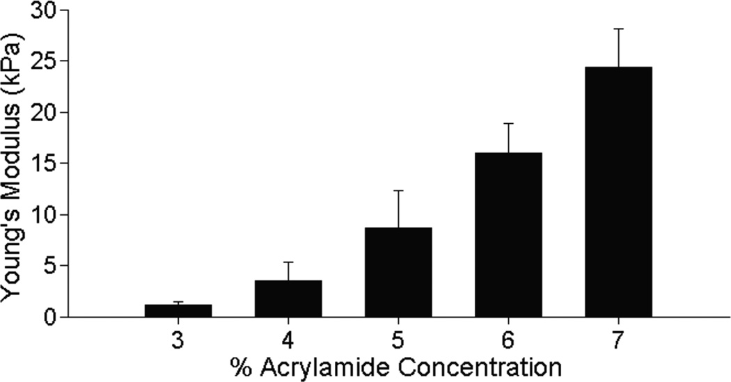
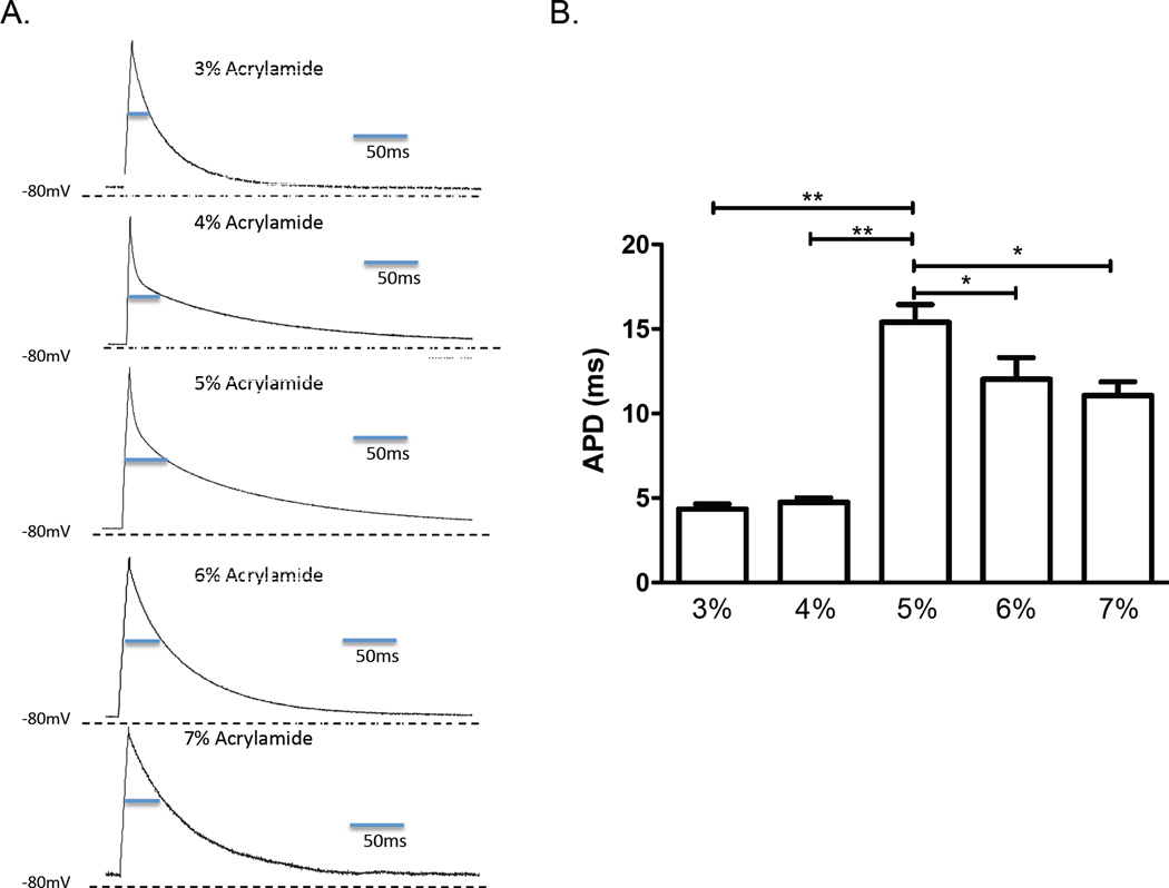
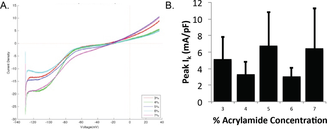
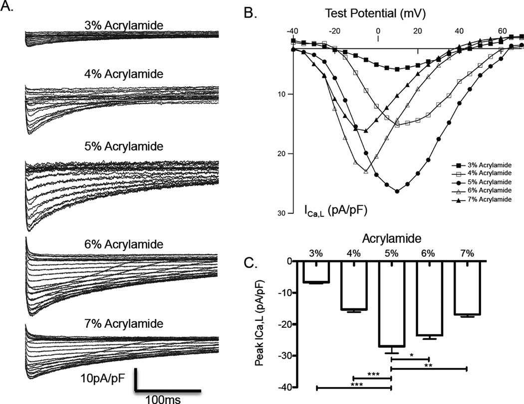
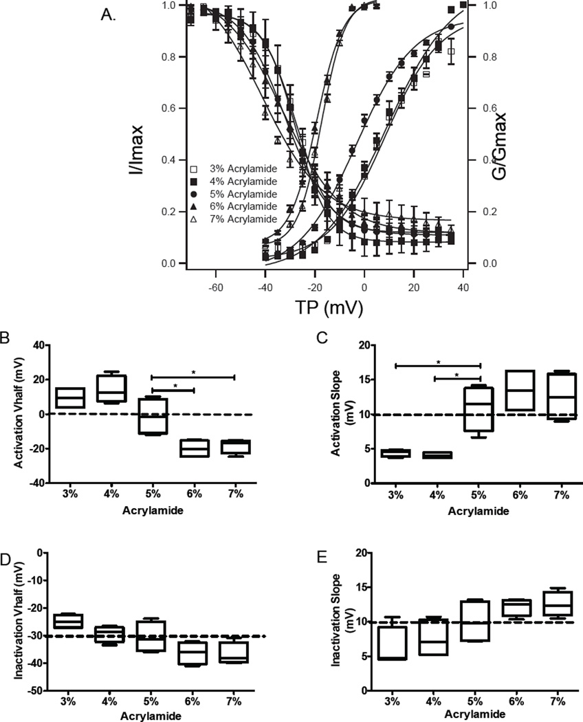
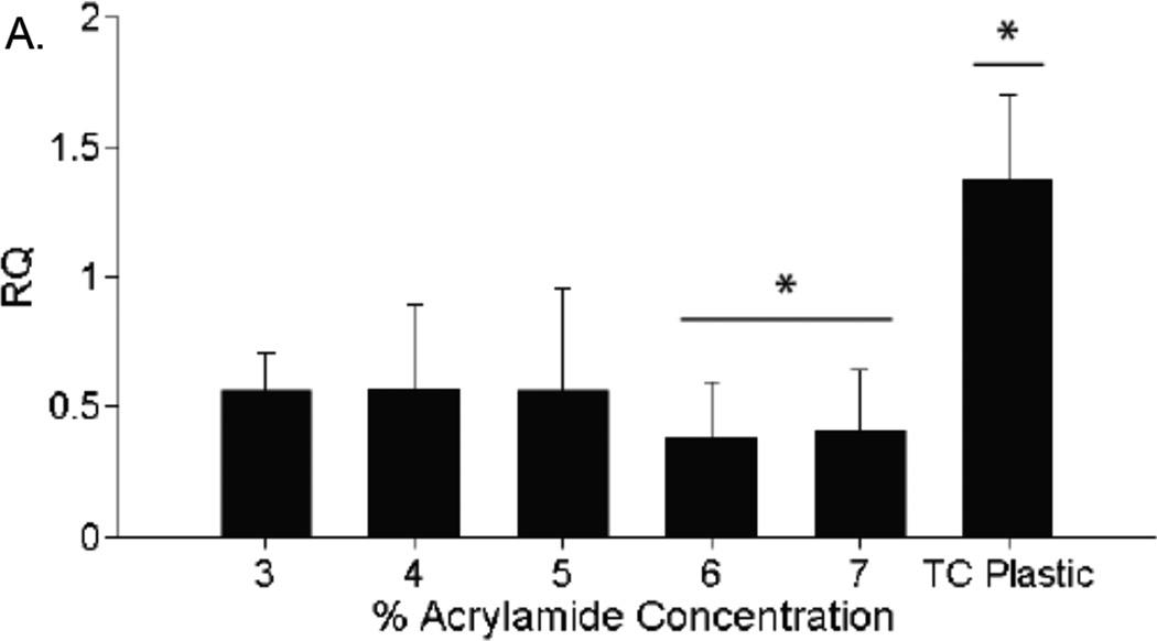
The stiffness of myocardial tissue changes significantly at birth and during neonatal development, concurrent with significant changes in contractile and electrical maturation of cardiomyocytes. Previous studies by our group have shown that cardiomyocytes generate maximum contractile force when cultured on a substrate with a stiffness approximating native cardiac tissue. However, effects of substrate stiffness on the electrophysiology and ion currents in cardiomyocytes have not been fully characterized. In this study, neonatal rat ventricular myocytes were cultured on the surface of flat polyacrylamide hydrogels with elastic moduli ranging from 1 to 25 kPa. Using whole-cell patch clamping, action potentials and L-type calcium currents were recorded. Cardiomyocytes cultured on hydrogels with a 9 kPa elastic modulus, similar to that of native myocardium, had the longest action potential duration. Additionally, the voltage at maximum calcium flux significantly decreased in cardiomyocytes on hydrogels with an elastic modulus higher than 9 kPa, and the mean inactivation voltage decreased with increasing stiffness. Interestingly, the expression of the L-type calcium channel subunit α gene and channel localization did not change with stiffness. Substrate stiffness significantly affects action potential length and calcium flux in cultured neonatal rat cardiomyocytes in a manner that may be unrelated to calcium channel expression. These results may explain functional differences in cardiomyocytes resulting from changes in the elastic modulus of the extracellular matrix, as observed during embryonic development, in ischemic regions of the heart after myocardial infarction, and during dilated cardiomyopathy.

Keywords: Calcium channels, L-type; Cardiac electrophysiology; Cardiac myoblasts; Elastic modulus; Patch clamp; Rats.


Figures

Figure 1
Figure 1
Elastic modulus of polyacrylamide hydrogels measured by imaging indentation depth of a steel ball bearing, and assuming Hertzian contact, shows that these hydrogels span the range of 1 to 25 kPa. n = 3 for all measurements.
Figure 2
Figure 2
Patch clamp-measured action potentials. A. Representative action potential traces of individual neonatal rat ventricular myocytes cultured for 7 days on hydrogels of varying elastic modulus. B. Composite data of action potential durations (APDs) at the 50th percentile (lines indicate significant differences with p* < 0.05 and p** < 0.01 compared to the APD on the 5% acrylamide gel; n = 5 for all reported values).
Figure 3
Figure 3
Patch voltage clamp-measured potassium currents. A. Representative potassium current traces of individual NRVM cultured for 7 days on hydrogels of varying elastic modulus; B. Peak potassium current (Ik) at holding potential of 20 mV on each hydrogel (no significant difference).
Figure 4
Figure 4
Patch voltage clamp-measured L-type calcium currents. A. Representative calcium current traces of individual NRVM cultured for 7 days on hydrogels of varying elastic modulus; B. Composite plot of voltage-mediated transmembrane L-type calcium currents (ICa,L) at holding potentials of −20 to 70mV. C. Peak calcium current on each hydrogel (p* < 0.05, p** < 0.01, p*** < 0.001). n = 4 for all reported values.
Figure 5
Figure 5
A. Steady state activation and inactivation of L-type calcium channels represented by the transmambrane calcium current normalized to maximum current (I/Imax) and the membrane conductance normalized to maximal conductance (G/Gmax). B–E. Half-voltage and slope for activation and inactivation curves.
Figure 6
Figure 6
Expression of L-type calcium channels. PCR results after amplifying RNA segments transcribed from the Cacna1c gene, which codes for the L-type calcium channel. Gene expression is not significantly different between any acrylamide gel groups. Gene expression in cells on polystyrene tissue culture plastic was significantly higher than in cells on acrylamide gels (p* < 0.05). n = 3 for all expression values.

References

    1. Jacot JG, Martin JC, Hunt DL. Mechanobiology of cardiomyocyte development. J Biomech. 2010;43:93–98. 2010. - PMC - PubMed
    1. Berry MF, Engler AJ, Woo YJ, Pirolli TJ, Bish LT, Jayasankar V, Morine KJ, Gardner TJ, Discher DE, Sweeney HL. Mesenchymal stem cell injection after myocardial infarction improves myocardial compliance. Am J Physiol Heart Circ Physiol. 2006;290:H2196–H2203. 2006. - PubMed
    1. Young JL, Engler AJ. Hydrogels with time-dependent material properties enhance cardiomyocyte differentiation in vitro. Biomaterials. 2011;32:1002–1009. 2011. - PMC - PubMed
    1. Jacot JG, McCulloch AD, Omens JH. Substrate stiffness affects the functional maturation of neonatal rat ventricular myocytes. Biophys J. 2008;95:3479–3487. 2008. - PMC - PubMed
    1. Tallawi M, Rai R, Boccaccini AR, Aifantis KE. Effect of substrate mechanics on cardiomyocyte maturation and growth. Tissue Eng Part B Rev. 2015;21:157–165. 2015. - PMC - PubMed

LinkOut - more resources