. 2016 Dec;17(1):35.
doi: 10.1186/s40510-016-0148-7. Epub 2016 Nov 7.

Craniofacial features as assessed by lateral cephalometric measurements in children with Down syndrome

Affiliations

Craniofacial features as assessed by lateral cephalometric measurements in children with Down syndrome

Veerasathpurush Allareddy et al. Prog Orthod. 2016 Dec.

Abstract

Objective: The objective of the present study is to examine the craniofacial development of patients with Down syndrome (DS) and compare them with a neurotypical population.

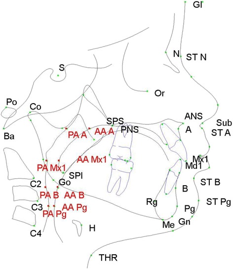
Methods: This study is a cross-sectional analysis of lateral cephalometric radiographs of participants with DS. The study population consisted of children and young adults with DS aged 3-25 years. Cephalometric data were summarized by age and sex. Raw and normalized z-scores were computed. One-sample t tests were used to test whether mean z-scores differed from zero. The demographic characteristics between those with or without lateral cephalograms among all study participants were compared by Fisher's exact tests.

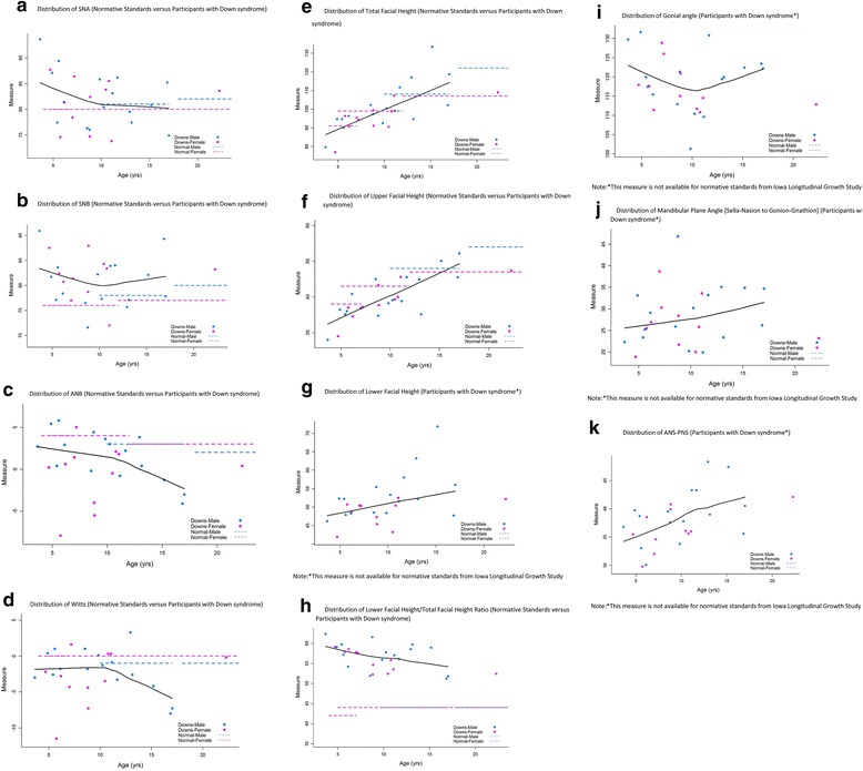
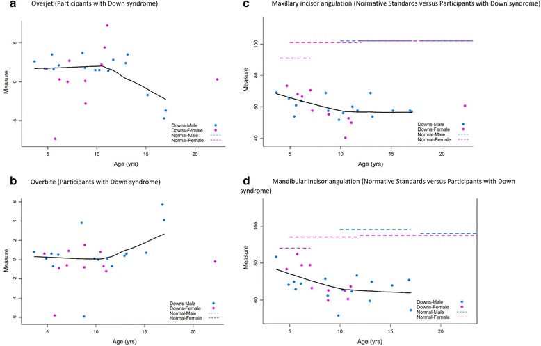
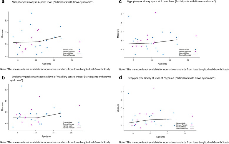
Results: The study sample comprised of 27 participants with DS. Study subjects demonstrated a class III skeletal pattern. This was more pronounced in the older age groups as compared to younger age groups. Subjects also had an increased proportionate lower anterior face height to total facial height compared to normative standards. Gonial angles, mandibular plane angles, and airway measurements increased with age.

Conclusions: Patients with Down syndrome present typically with class III skeletal pattern and long lower anterior facial heights. In patients with Down syndrome, comprehensive phase of orthodontic treatment may be best initiated following cessation of growth.


Figures

Fig. 1
Fig. 1
Diagrammatic representation of anatomic landmarks used to identify craniofacial and soft tissue parameters on cephalometric radiographs. Sixty one landmarks and 40 measurements were included in the custom analysis. Airway landmarks are highlighted in red. See Table 1 for explanation of abbreviations
Fig. 2
Fig. 2
Skeletal maxillary, mandibular, and inter-maxillary measurements. a Distribution of SNA (normative standards versus participants with Down syndrome). b Distribution of SNB (normative standards versus participants with Down syndrome). c Distribution of ANB (normative standards versus participants with Down syndrome). d Distribution of Wits (normative standards versus participants with Down syndrome). e Distribution of total facial height (normative standards versus participants with Down syndrome). f Distribution of upper facial height (normative standards versus participants with Down syndrome). g Distribution of lower facial height (participants with Down syndrome*). h Distribution of lower facial height to total facial height ratio (normative standards versus participants with Down syndrome). i Distribution of gonial angle (participants with Down syndrome*). j Distribution of mandibular plane angle [sella-nasion to gonion-gnathion] (participants with Down syndrome*). k Distribution of ANS-PNS (participants with Down syndrome*)
Fig. 3
Fig. 3
Overjet, overbite, and incisor angulations. a Overjet (participants with Down syndrome). b. Overbite (participants with Down syndrome). c. Maxillary incisor angulation (normative standards versus participants with Down syndrome). d. Mandibular incisor angulation (normative standards versus participants with Down syndrome)
Fig. 4
Fig. 4
Airway Measurements. a. Nasopharynx airway at A point level (participants with Down syndrome*). b. Oral pharyngeal airway space at level of maxillary central incisor (participants with Down syndrome*). c. Hypopharynx airway space at B point level (participants with Down syndrome*). d. Deep pharynx airway at level of pogonion (participants with Down syndrome*)

References

    1. Gorlin RJ, Cohen MM, Levin LS. Syndromes of the head and neck. 4. New York, NY: Oxford University Press; 2001. pp. 35–42.
    1. de Graaf G, Buckley F, Skotko BG. Estimates of the live births, natural losses, and elective terminations with Down syndrome in the United States. Am J Med Genet Part A. 2015;167A(4):756–67. doi: 10.1002/ajmg.a.37001. - DOI - PubMed
    1. Stoll C, Alembik Y, Dott B, Roth MP. Study of Down syndrome in 238,942 consecutive births. Ann Genet. 1998;41(1):44–51. - PubMed
    1. Silva Jesuino FA, Valladares-Neto J. Craniofacial morphological differences between Down syndrome and maxillary deficiency children. Eur J Orthod. 2013;35(1):124–30. doi: 10.1093/ejo/cjr105. - DOI - PubMed
    1. Suri S, Tompson BD, Cornfoot L. Cranial base, maxillary and mandibular morphology in Down syndrome. Angle Orthodontist. 2010. Vol 80, No 5. - PMC - PubMed

MeSH terms

LinkOut - more resources