. 2017 Apr 3;11(2):112-120.
doi: 10.1080/19336934.2016.1244591. Epub 2016 Oct 10.

Successive requirement of Glass and Hazy for photoreceptor specification and maintenance in Drosophila

Affiliations

Successive requirement of Glass and Hazy for photoreceptor specification and maintenance in Drosophila

F Javier Bernardo-Garcia et al. Fly (Austin). .

Abstract

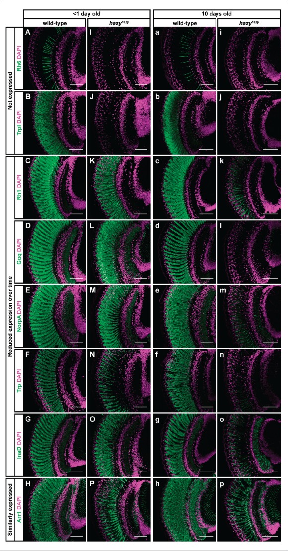
Development of the insect compound eye requires a highly controlled interplay between transcription factors. However, the genetic mechanisms that link early eye field specification to photoreceptor terminal differentiation and fate maintenance remain largely unknown. Here, we decipher the function of 2 transcription factors, Glass and Hazy, which play a central role during photoreceptor development. The regulatory interactions between Glass and Hazy suggest that they function together in a coherent feed-forward loop in all types of Drosophila photoreceptors. While the glass mutant eye lacks the expression of virtually all photoreceptor genes, young hazy mutants correctly express most phototransduction genes. Interestingly, the expression of these genes is drastically reduced in old hazy mutants. This age-dependent loss of the phototransduction cascade correlates with a loss of phototaxis in old hazy mutant flies. We conclude that Glass can either directly or indirectly initiate the expression of most phototransduction proteins in a Hazy-independent manner, and that Hazy is mainly required for the maintenance of functional photoreceptors in adult flies.

Keywords: Drosophila; Glass; Hazy; Pph13; cell fate maintenance; eye development; photoreceptor differentiation; phototransduction.


Figures

Figure 1.
Figure 1.
Expression analysis of the hazy(wt)-GFP reporter in the ocelli and Bolwig's organ PRs. (A-C) In the case of the ocelli, these are 3 visual organs located dorsally on the head of adult flies (A). Samples were stained with antibodies against GFP (green), and Elav (used as a neuronal marker, magenta). The hazy(wt)-GFP reporter was expressed in the ocelli in wild-type (A, A'), but not glass mutant background (B). A hazy(gl1,2mut)-GFP reporter in which the 2 Glass binding sites were mutated was not expressed in the ocelli (C). (D-F) In the case of the Bolwig's organ, this is a larval eye that develops from the optic placode during embryogenesis. Stage 14 embryos were stained with antibodies against GFP (green), Fas2 (red) and Kruppel (blue). At this stage, the developing Bolwig's organ is located dorsally, still in contact with the surface, and can be identified both because of its position and the co-expression of Fas2 and Kruppel.,, Similar to the ocelli, the hazy(wt)-GFP reporter was expressed in the Bolwig's organ in wild-type (D), but not glass mutant background (E). Also, hazy(gl1,2mut)-GFP was not expressed in the Bolwig's organ (F). For each image, the 3 channels from a close-up of the Bolwig's organ were separated and are shown below in grayscale (D′-F‴). Scale bars represent 20 μm in D′-F‴; 30 μm in A′, B-F; and 100 μm in A.
Figure 2.
Figure 2.
Test for additional regulatory interactions between Glass and Hazy. (A, B) We used Hoechst (blue), which labels cell nuclei, as a counterstain to analyze the expression pattern of the glass-DsRed and hazy(wt)-GFP reporters in the CNS of third instar larvae. The glass-DsRed reporter was expressed the nuclei of some cells in the brain (green, A). A close-up to the right shows that those neurons endogenously expressing Glass (red/magenta) also co-express the reporter (green, A′). These two channels are shown separately to the right in grayscale (A,″ A‴). The hazy(wt)-GFP reporter is exclusively expressed in PRs (green, B). A grayscale image to the right shows GFP labeling the axonal projections of the PRs in the brain (arrows, B′). (C-E) In flip-out experiments we ectopically induced either Glass or Hazy expression in clones labeled with nuclear β-galactosidase (βGal). We stained the CNS of third instar larvae with antibodies against βGal (red/magenta); either DsRed, GFP or Glass (green) and with Hoechst (blue). We found that Glass ectopically induced the expression of the glass-DsRed reporter in the ventral nerve cord (C, C′; channels are also shown separately in grayscale in C,″ C‴). By contrast, Hazy did not ectopically induce the hazy(wt)-GFP reporter (D, D′) nor Glass (E, E′). Scale bars represent 20 μm in A′, C′- E′; and 80 μm in A-E.
Figure 3.
Figure 3.
Expression of phototransduction proteins in the hazy mutant retina. Head sections were taken of control and hazyhazy flies, and stained with antibodies against different phototransduction proteins (green) and with Hoechst (used to label cell nuclei, magenta). (A-P) One group of flies was dissected on the day they eclosed. At this age we did not detect neither Rh6 (I) nor Trpl (J) in the retina of hazy mutants, but most of the phototransduction proteins that we tested were correctly expressed: Rh1 (K), Gαq (L), NorpA (M), Trp (N), InaD (O) and Arr1 (P). (a-p) A second group of flies were dissected 10 d after eclosion. Neither Rh6 (i) nor Trpl (j) were expressed in the retina of these older hazy mutants, and most phototransduction proteins showed decreased expression levels: Rh1 (k), Gαq (l), NorpA (m), Trp (n) and InaD (o). Only Arr1 (p) expression did not seem reduced over time in the hazy mutant retina. Scale bars represent 50 µm.
Figure 4.
Figure 4.
Age-related changes in the phototaxis of wild-type, glass and hazy mutant flies. Box plots show the light preference indices (PIs) of wild-type (yellow), glass (cyan) and hazy mutants (pink) of different ages. Bold lines represent medians. The upper and lower quartiles are represented by the top and the bottom of each box. Whisker lines indicate the maximum and minimum data point that are closer than 1.5 interquartile range of its nearest quartile. Circles indicate outliers. We used Welch's t-test for comparing the PIs between groups (n = 7 per age and genotype) and to zero. Significance levels represent p > 0.05 (not significant, n.s.), p ≤ 0.05 (*), p ≤ 0.01 (**), and p ≤ 0.001 (***). In a 2-choice assay, groups of wild-type flies of every age showed positive phototaxis, which decreases with age (indicated by positive PI values, which were significantly different from zero). glass mutants were photoneutral at all ages (their PIs were not significantly different from zero). Newly eclosed hazy mutants showed positive phototaxis, not different from that of wild-type flies (p = 0.67, median wild-type PI = 0.83). Five day old hazy mutants and wild-type flies show a decreased positive phototaxis, but their PIs are not different from each other (p = 0.30, median wild-type PI = 0.42). Ten day old hazy mutants were photoneutral, with their PIs comparable to zero (p = 0.08) or to glass mutants (p = 0.56), and significantly different from wild-type (median wild-type PI = 0.22).

Comment on

  • Extra View to: Bernardo-Garcia F. J., Fritsch C., Sprecher S. G. The transcription factor Glass links eye field specification with photoreceptor differentiation in Drosophila. Development 2016; 143:1413-1423.

References

    1. Tsachaki M, Sprecher SG. Genetic and developmental mechanisms underlying the formation of the Drosophila compound eye. Dev Dyn 2012; 241:40-56; PMID:21932322; http://dx.doi.org/10.1002/dvdy.22738 - DOI - PubMed
    1. Treisman JE. Retinal differentiation in Drosophila. Wiley Interdiscip Rev Dev Biol 2013; 2:545-57; PMID:24014422; http://dx.doi.org/10.1002/wdev.100 - DOI - PMC - PubMed
    1. Potier D, Davie K, Hulselmans G, Naval Sanchez M, Haagen L, Huynh-Thu VA, Koldere D, Celik A, Geurts P, Christiaens V, et al.. Mapping gene regulatory networks in Drosophila eye development by large-scale transcriptome perturbations and motif inference. Cell Rep 2014; 9:2290-303; PMID:25533349; http://dx.doi.org/10.1016/j.celrep.2014.11.038 - DOI - PubMed
    1. Halder G, Callaerts P, Gehring WJ. Induction of ectopic eyes by targeted expression of the eyeless gene in Drosophila. Science 1995; 267:1788-92; PMID:7892602; http://dx.doi.org/10.1126/science.7892602 - DOI - PubMed
    1. Pignoni F, Hu B, Zavitz KH, Xiao J, Garrity PA, Zipursky SL. The eye-specification proteins So and Eya form a complex and regulate multiple steps in Drosophila eye development. Cell 1997; 91:881-91; PMID:9428512; http://dx.doi.org/10.1016/S0092-8674(00)80480-8 - DOI - PubMed

MeSH terms

LinkOut - more resources