. 2017 Feb;16(2):230-235.
doi: 10.1038/nmat4772. Epub 2016 Oct 10.

Single-platelet nanomechanics measured by high-throughput cytometry

Affiliations

Single-platelet nanomechanics measured by high-throughput cytometry

David R Myers et al. Nat Mater. 2017 Feb.

Abstract

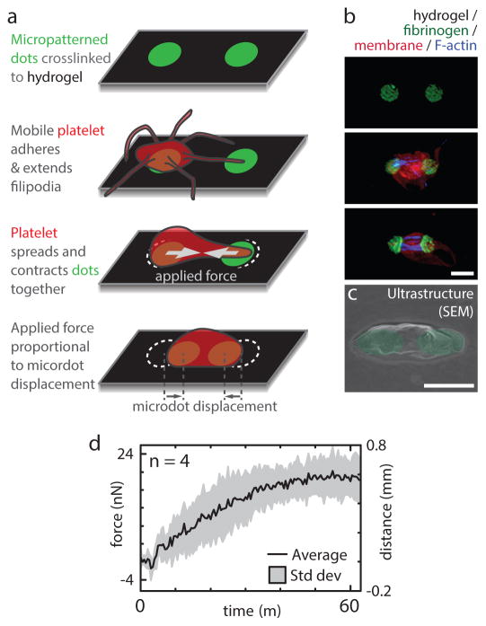
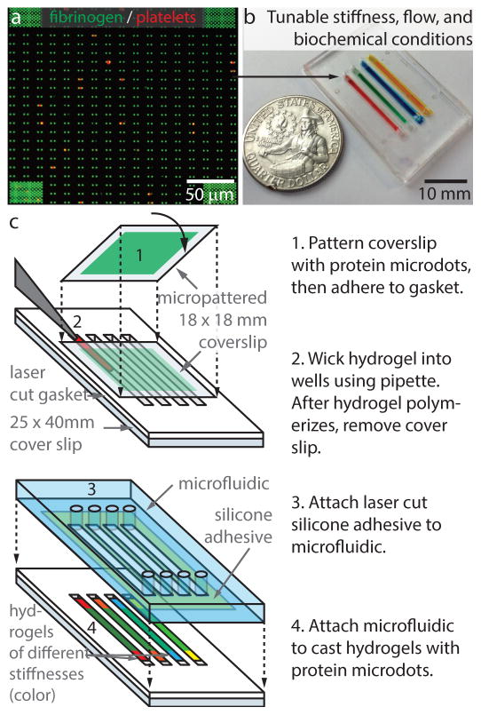
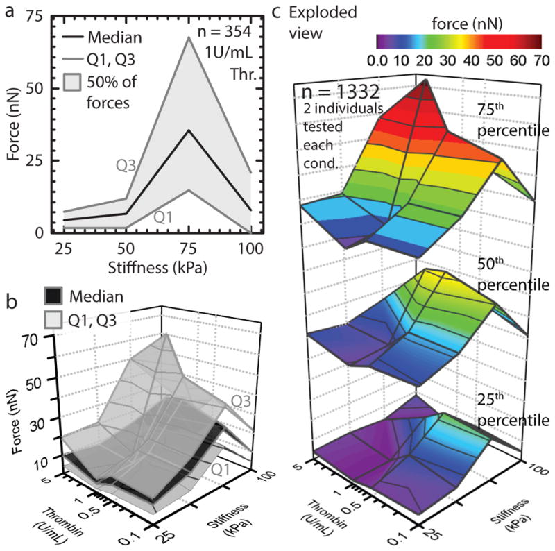
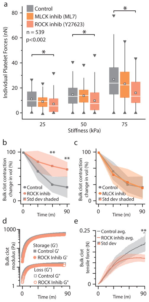
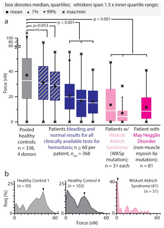
Haemostasis occurs at sites of vascular injury, where flowing blood forms a clot, a dynamic and heterogeneous fibrin-based biomaterial. Paramount in the clot's capability to stem haemorrhage are its changing mechanical properties, the major drivers of which are the contractile forces exerted by platelets against the fibrin scaffold. However, how platelets transduce microenvironmental cues to mediate contraction and alter clot mechanics is unknown. This is clinically relevant, as overly softened and stiffened clots are associated with bleeding and thrombotic disorders. Here, we report a high-throughput hydrogel-based platelet-contraction cytometer that quantifies single-platelet contraction forces in different clot microenvironments. We also show that platelets, via the Rho/ROCK pathway, synergistically couple mechanical and biochemical inputs to mediate contraction. Moreover, highly contractile platelet subpopulations present in healthy controls are conspicuously absent in a subset of patients with undiagnosed bleeding disorders, and therefore may function as a clinical diagnostic biophysical biomarker.


Conflict of interest statement

The authors declare no competing financial interests.

Figures

Figure 1
Figure 1
The fundamental unit driving clot stiffening, a single platelet pulling against a fibrin/ogen substrate, is established by recapitulating the mechanical and biological microenvironment of the platelet. a, Fluorescently-conjugated fibrinogen microdot pairs are covalently bound to a deformable polyacrylamide hydrogel of known mechanical properties. As a platelet adheres and pulls pairs of fibrinogen microdots together, the contractile force is proportional to the microdot displacement. b, Super-resolution microscopy images of individual platelets with various degrees of contraction, scale bar is 2 μm c, Scanning electron microscopy image of a platelet contracting a fibrinogen microdot pair, scale bar is 2 μm. d, Real time contractile measurements of 4 single platelets.
Figure 2
Figure 2
Platelet contraction cytometer - hydrogels with microprinted arrays of fibrinogen microdots are encapsulated in separate microchannels, enabling the biochemical, mechanical, and shear microenvironments to be precisely controlled and varied simultaneously. a, A confocal image showing single platelets (red, contracting against fibrinogen microdot pairs (green) on the hydrogel surface. Over 20,000 fibrinogen microdot pairs are microprinted on the surface of each hydrogel encapsulated in each microchannel. b, Each microfluidic device may comprise different variables,, here four of microchannels comprise of hydrogels of different stiffness. c, A novel yet relatively simple fabrication process flow enables rapid manufacturing.
Figure 3
Figure 3
Biochemical and mechanical cues synergistically mediate platelet contraction force. a, Maximal platelet contraction occurs at 75kpa substrate stiffness and 1U/mL thrombin. b, Single platelet force quartiles for physiologically relevant clot stiffnesses and thrombin concentrations. Platelet contractile forces are highest at a substrate stiffness of 75 kPa or at 5U/mL thrombin. A minimum occurs at 25 kPa stiffness and 1U/mL thrombin. c, Exploded view of b to aid visualization. Microenvironmental stiffness has a dominant role over thrombin concentration. Force attenuation at the highest stiffness conditions suggests a mechanically mediated negative feedback mechanism governing the upper limits of platelet contraction. n refers to total number of platelets tested.
Figure 4
Figure 4
Mechanotransductive platelet contraction is mediated by the Rho-associated protein kinase (ROCK) pathway, as measured with platelet contraction cytometry and standard bulk clot contraction and bulk clot rheology. a, The increase in platelet contractile forces with increasing substrate stiffness is significantly reduced with exposure to Y27623, a pharmacologic ROCK inhibitor. Pharmacologic inhibition of the myosin light chain kinase (MLCK) with ML7, on the other hand, did not produce a statistically significant difference in the substrate stiffness-mediated effect on platelet contractile force. Box denotes median and quartiles; whiskers to 1.5 interquartlie range), square denotes median, triangles denotes 1% and 99%. (n = 539, each condition n>40). * indicates differences from control at same stiffness (p < 0.05 by Mann-Whitney). b, ROCK inhibition impairs bulk clot contraction (n = 4) as compared to the untreated control clots. c, MLCK inhibition does not change bulk clot contraction (n = 4). d, Oscillatory rheology, which enables simultaneous measurements of storage (G′) and loss (G″) moduli as well as bulk tensile forces, revealed that both control and ROCK-inhibited clots undergo a dramatic stiffening process (representative plot from 3 similar experiments shown). The angular frequency and strain amplitude are 1 rad/s and 0.01, respectively, which are well within the linear regime of the samples. e, Control and ROCK-inhibited clots apply similar forces during the beginning of clot formation when the measured storage and loss moduli are low (n = 3). As the clots stiffen (Figure 4d), ROCK-inhibited bulk clots begin to exert lower tensile forces and plateau while the control continues to increase over time. ** indicates difference from control (p < 0.05 by t-test).
Figure 5
Figure 5
Patients with phenotypic bleeding lack highly contractile platelets associated with clot contraction and force generation. a, Wiskott Aldrich Syndrome and May Hegglin Disorder platelets exhibited significantly reduced contractile forces compared to that of healthy controls (75 kPa gel stiffness, 1U/mL thrombin). In a subset of patients with bleeding diatheses yet normal hemostasis tests, platelet contraction was lower than that of normal healthy controls. b, Histogram data reveal platelet subpopulations of varying contractile forces. Healthy control platelets comprise high contractility subpopulations, notably absent in platelets from a Wiskott Aldrich Syndrome patient. In all panels, n refers to number of platelets, Mann-Whitney statistical test.

References

    1. Jen CJ, McIntire LV. The structural properties and contractile force of a clot. Cell Motil. 1982;2:445–55. - PubMed
    1. HVAS & Sørensen. Tranexamic acid combined with recombinant factor VIII increases clot resistance to accelerated fibrinolysis in severe hemophilia A. 2007 doi: 10.1111/j.1538-7836.2007.02755.x. - DOI - PubMed
    1. Collet JP, et al. Altered fibrin architecture is associated with hypofibrinolysis and premature coronary atherothrombosis. Arterioscler Thromb Vasc Biol. 2006;26:2567–73. - PubMed
    1. Carr ME. Development of platelet contractile force as a research and clinical measure of platelet function. Cell Biochem Biophys. 2003;38:55–78. - PubMed
    1. Cohen I, De Vries A. Platelet contractile regulation in an isometric system. Nature. 1973;246:36–7. - PubMed

Publication types

MeSH terms

LinkOut - more resources