

doi: 10.1038/nchembio.2180.
Epub 2016 Oct 10.
Serine is a new target residue for endogenous ADP-ribosylation on histones
Affiliations
- PMID: 27723750
- PMCID: PMC5113755
- DOI: 10.1038/nchembio.2180
Item in Clipboard
Serine is a new target residue for endogenous ADP-ribosylation on histones
Nat Chem Biol.
2016 Dec.
Abstract
ADP-ribosylation (ADPr) is a biologically and clinically important post-translational modification, but little is known about the amino acids it targets on cellular proteins. Here we present a proteomic approach for direct in vivo identification and quantification of ADPr sites on histones. We have identified 12 unique ADPr sites in human osteosarcoma cells and report serine ADPr as a new type of histone mark that responds to DNA damage.
Conflict of interest statement
Statement The authors declare no competing financial interests.
Figures
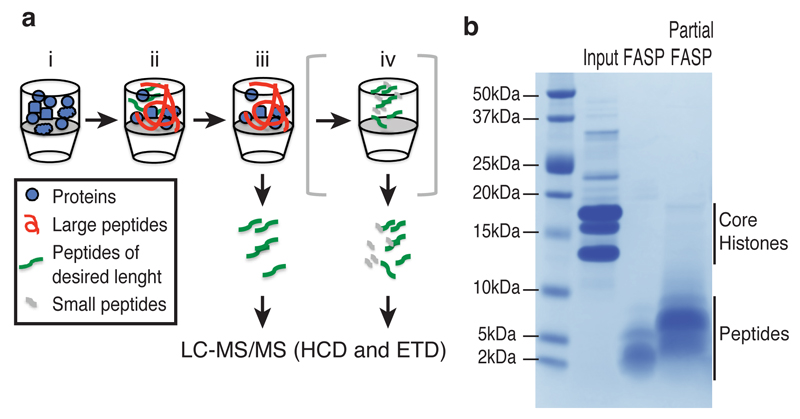
(a) Workflow of the Partial FASP method. Histones are digested on the filter with limiting amount of trypsin (1:2000) for 20 minutes at 20 ºC (i, ii), the peptides amenable for MS analysis are separated from the undigested proteins by centrifugation (iii). Optionally, undigested proteins and large peptides can be further digested overnight to recover smaller peptides similarly to the FASP method (iv). See Online Methods for further details. (b) SDS-PAGE analysis of the purified histones (Input, 10 μg) and eluted peptides from the filter after a full digestion (FASP) or a partial digestion (Partial FASP) of 100 μg proteins. See Supplementary Fig. 10 for original image.
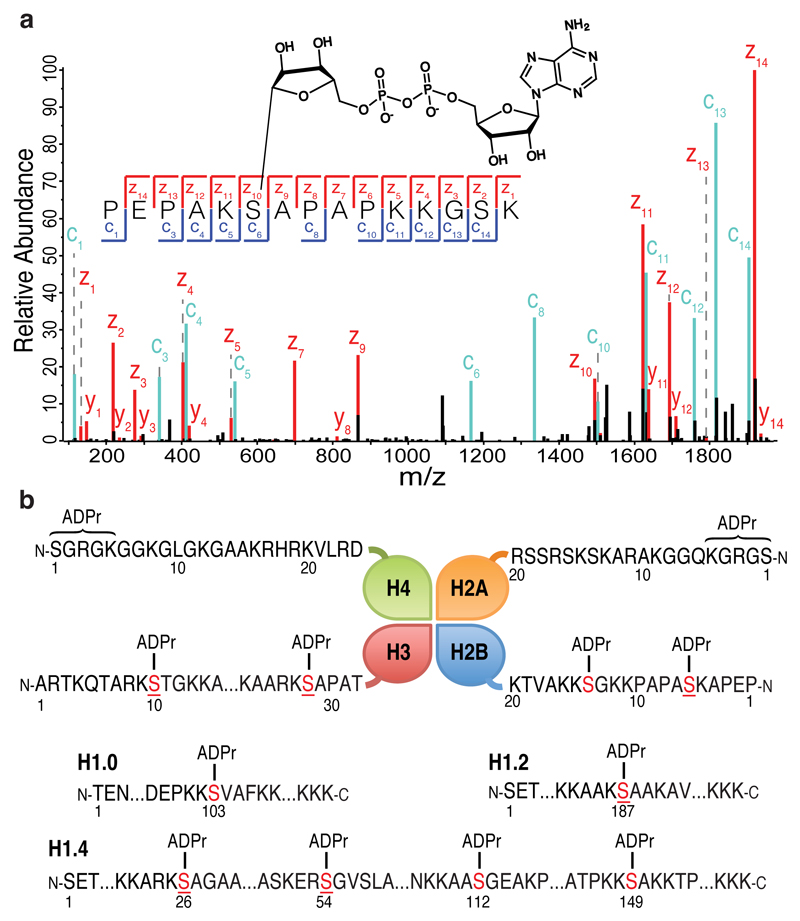
(a) High-resolution ETD fragmentation spectra of an H2B peptide modified by ADP-ribose on Serine 6. The chemical structure of ADP-ribose is depicted. See Supplementary Fig. 7a for further details on the linkage between Serine and ADPr. (b) Schematic representation of core histones (top panel) and different H1 variants (bottom panel). The twelve novel unique ADPr marks are indicated. ADPr serine residues (in red) are located on the N-terminal tails of the four core histones. Underlined serines are reported phosphorylation sites.
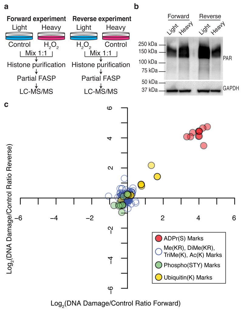
(a) Schematic representation of the SILAC-based strategy to quantify core histone marks upon 10 minutes of 2 mM H2O2-induced DNA damage. (b) Western blot analysis of total protein poly-ADP-ribosylation levels prior to mixing light and heavy lysates from each SILAC experiment. Anti-GAPDH was used as a loading control. See Supplementary Fig. 10 for original images. (c) Log transformed Heavy/Light SILAC ratios from the forward and Light/Heavy SILAC ratios from the reverse experiment were plotted against each other. Each point represents a unique histone mark.
References
-
- Hottiger MO. Nuclear ADP-Ribosylation and Its Role in Chromatin Plasticity, Cell Differentiation, and Epigenetics. Annu Rev Biochem. 2015;84:227–263. - PubMed
MeSH terms
Substances
Grants and funding
LinkOut - more resources
Full Text Sources
Other Literature Sources
Molecular Biology Databases
