. 2016 Dec;111(12):1726-1735.
doi: 10.1038/ajg.2016.454. Epub 2016 Oct 11.

Evaluation of Esophageal Motility Utilizing the Functional Lumen Imaging Probe

Affiliations

Evaluation of Esophageal Motility Utilizing the Functional Lumen Imaging Probe

Dustin A Carlson et al. Am J Gastroenterol. 2016 Dec.

Abstract

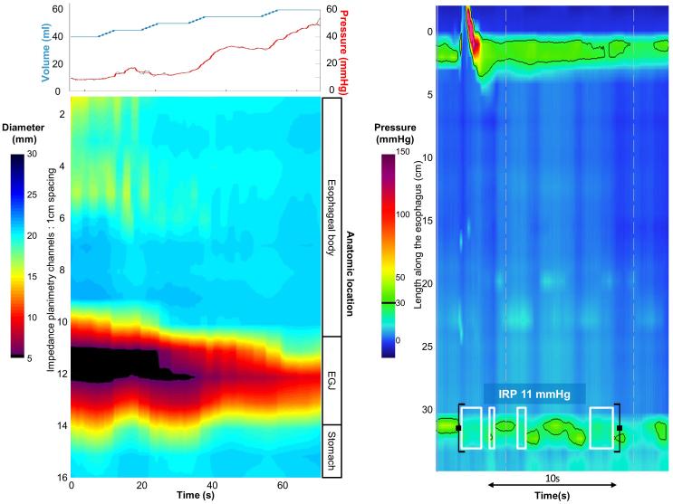
Objectives: Esophagogastric junction (EGJ) distensibility and distension-mediated peristalsis can be assessed with the functional lumen imaging probe (FLIP) during a sedated upper endoscopy. We aimed to describe esophageal motility assessment using FLIP topography in patients presenting with dysphagia.

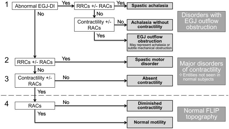
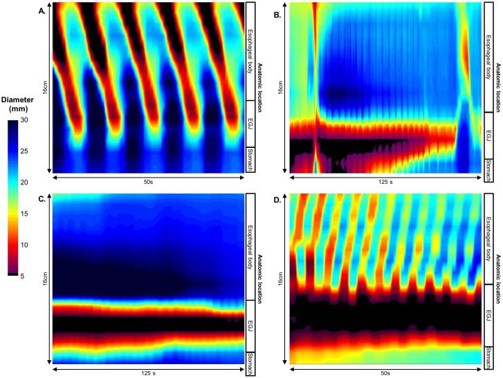
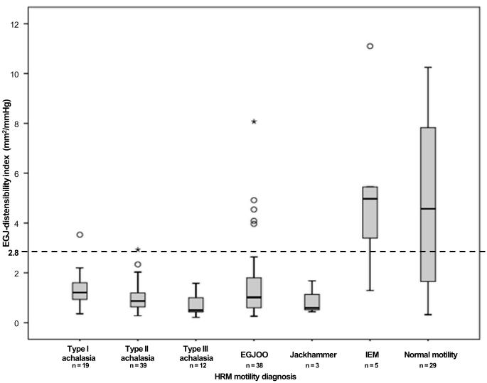
Methods: In all, 145 patients (aged 18-85 years, 54% female) with dysphagia that completed upper endoscopy with a 16-cm FLIP assembly and high-resolution manometry (HRM) were included. HRM was analyzed according to the Chicago Classification of esophageal motility disorders; major esophageal motility disorders were considered "abnormal". FLIP studies were analyzed using a customized program to calculate the EGJ-distensibility index (DI) and generate FLIP topography plots to identify esophageal contractility patterns. FLIP topography was considered "abnormal" if EGJ-DI was <2.8 mm2/mm Hg or contractility pattern demonstrated absent contractility or repetitive, retrograde contractions.

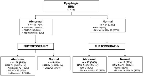
Results: HRM was abnormal in 111 (77%) patients: 70 achalasia (19 type I, 39 type II, and 12 type III), 38 EGJ outflow obstruction, and three jackhammer esophagus. FLIP topography was abnormal in 106 (95%) of these patients, including all 70 achalasia patients. HRM was "normal" in 34 (23%) patients: five ineffective esophageal motility and 29 normal motility. In all, 17 (50%) had abnormal FLIP topography including 13 (37%) with abnormal EGJ-DI.

Conclusions: FLIP topography provides a well-tolerated method for esophageal motility assessment (especially to identify achalasia) at the time of upper endoscopy. FLIP topography findings that are discordant with HRM may indicate otherwise undetected abnormalities of esophageal function, thus FLIP provides an alternative and complementary method to HRM for evaluation of non-obstructive dysphagia.


Figures

Figure 1
Figure 1. FLIP topography motility classification
FLIP topography motility diagnoses were generated using a hierarchical analysis scheme based on 1) EGJ-distensibility index (DI) and 2) Contractile pattern. Motility diagnoses above the dashed line represent patterns not observed in asymptomatic controls and therefore considered major motor disorders, i.e. ‘abnormal’ FLIP topography. RACs – repetitive, antegrade contractions. RRCs – repetitive, retrograde contractions.
Figure 2
Figure 2. FLIP topography contractile patterns.
A) Repetitive, antegrade contractions (RACs) from a patient with normal motility on high-resolution manometry (HRM). RACs were the most commonly observed contractile pattern in asymptomatic controls.(10, 11) B) Contractility without RAC or repetitive, retrograde contractions in a patients with a HRM demonstrating EGJ outflow obstruction with a peristaltic pattern of ineffective esophageal motility. C) Absent contractility in a patient with type I achalasia on HRM. D). Repetitive, retrograde contractions (RRCs) in a patient with type III achalasia on HRM.
Figure 3
Figure 3. EGJ – distensibility index by manometric esophageal motility diagnosis
The dashed line indicates the lower range of normal based on previous study of asymptomatic controls.(10, 11) EGJOO – esophagogastric junction outflow obstruction. IEM – ineffective esophageal motility. HRM – high resolution manometry.
Figure 4
Figure 4. Flow diagram of high-resolution manometry (HRM) and FLIP topography motility diagnoses
“Abnormal” indicated detection of a major esophageal motility disorder. DX – diagnosis. EGJOO – EGJ outflow obstruction. IEM – ineffective esophageal motility.
Figure 5
Figure 5. Case examples of FLIP topography
Left panels represent portions of the FLIP study: distension volume (top, blue line), and intra-balloon pressure (top, red line), and FLIP topography (bottom). A swallow from the corresponding HRM is included in the right panels. A) A patient with type I achalasia, but borderline IRP (median 12 mmHg). FLIP topography demonstrated an abnormal EGJ-DI of 1.6 mm2/mmHg and absent contractility. B) A patient with EGJOO, and suspected evolving achalasia, on HRM. FLIP topography demonstrated abnormal EGJ-DI and absent contractility, supporting the diagnosis of achalasia. The patients was treated with a botulinum-toxin injection to the lower esophageal sphincter that resulted in improvement in dysphagia. C) A patient with EGJOO on HRM. FLIP topography was essentially normal with a normal EGJ-DI (4.9 mm2/mmHg) and RACs. An esophagram showed normal clearance of barium. Dysphagia had resolved without intervention at 6-month follow-up. D) A patient with normal motility on HRM (median IRP 13 mmHg), but with an abnormal EGJ-DI (0.46 mm2/mmHg) and RRCs on FLIP topography. Esophagram demonstrated persistent esophageal barium column height of 4.4-cm after 5 minutes and impaction of a 12.5 mm barium tablet at the EGJ. The patient underwent endoscopic ultrasound with diffusely thickened distal esophageal muscle layers supporting diagnosis of a primary esophageal motor disorder. A per-oral endoscopic myotomy was recommended

References

    1. Xiao Y, Kahrilas PJ, Nicodeme F, et al. Lack of correlation between HRM metrics and symptoms during the manometric protocol. Am J Gastroenterol. 2014;109(4):521–6. - PMC - PubMed
    1. Rohof WO, Hirsch DP, Kessing BF, et al. Efficacy of treatment for patients with achalasia depends on the distensibility of the esophagogastric junction. Gastroenterology. 2012;143(2):328–35. - PubMed
    1. Pandolfino JE, de Ruigh A, Nicodeme F, et al. Distensibility of the esophagogastric junction assessed with the functional lumen imaging probe (FLIP) in achalasia patients. Neurogastroenterol Motil. 2013;25(6):496–501. - PMC - PubMed
    1. Smeets FG, Masclee AA, Keszthelyi D, et al. Esophagogastric junction distensibility in the management of achalasia patients: relation to treatment outcome. Neurogastroenterol Motil. 2015;27(10):1495–503. - PubMed
    1. Orvar KB, Gregersen H, Christensen J. Biomechanical characteristics of the human esophagus. Dig Dis Sci. 1993;38(2):197–205. - PubMed

Publication types

MeSH terms