. 2016 Sep 1;6(9):1935-1948.
eCollection 2016.

Identification of HRAS as cancer-promoting gene in gastric carcinoma cell aggressiveness

Affiliations

Identification of HRAS as cancer-promoting gene in gastric carcinoma cell aggressiveness

Xiao Yu Wu et al. Am J Cancer Res. .

Abstract

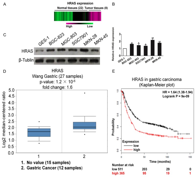
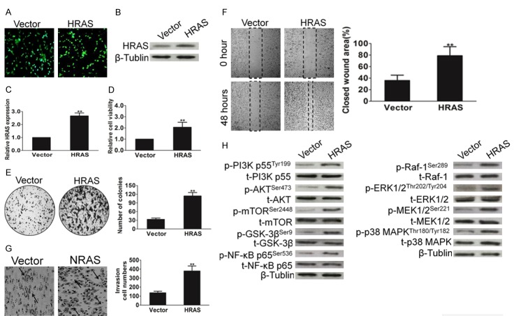
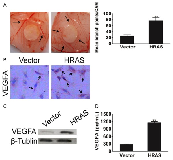
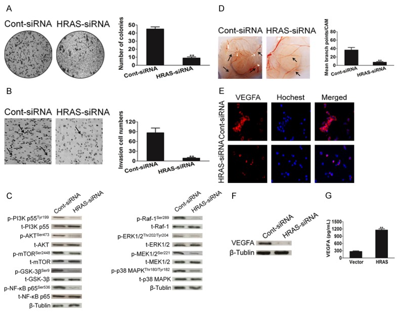
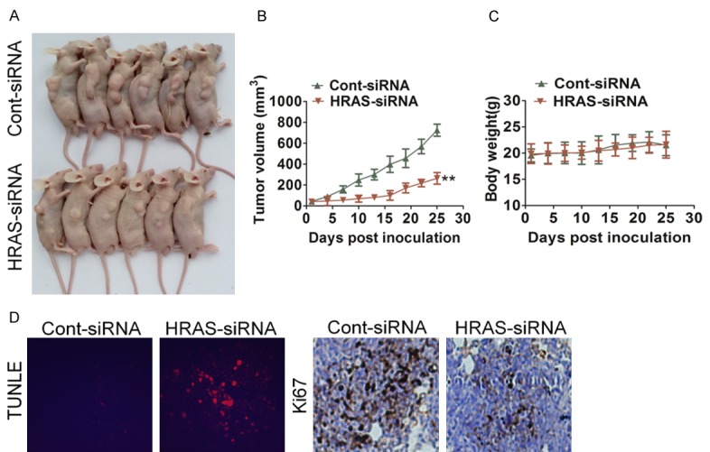
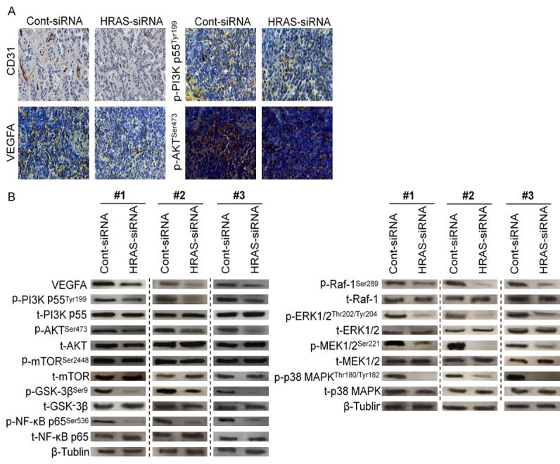
Gastric carcinoma is one of the most lethal malignancies of cancers and its prognosis remains dismal due to the paucity of effective therapeutic targets. Herein, we showed that HRAS is markedly up-regulated in gastric carcinoma. Prognostic analysis indicated that HRAS expression might be a prognostic indicator for the survival of patients with gastric carcinoma. Ectopic expression of HRAS in gastric carcinoma cells accelerated proliferation, migration, invasion, angiogenesis, and clone formation ability of gastric carcinoma cells in vitro. Furthermore, HRAS over-expressing significantly promoted the tumorigenicity of gastric carcinoma cells in vivo whereas silencing endogenous HRAS caused opposite outcomes. Moreover, we demonstrated that HRAS enhanced gastric carcinoma aggressiveness by activating VEGFA/PI3K/AKT pathway and Raf-1 signaling. Together, our results provide new evidence that HRAS overexpression promotes the progression of gastric carcinoma and might represent a novel therapeutic target for its treatment.

Keywords: HRAS; angiogenesis; gastric carcinoma; growth.


Figures

Figure 1
Figure 1
Overexpression of HRAS correlates with poor prognosis of gastric carcinoma. A. Expression profiling of mRNAs showing that HRAS was up-regulated in gastric carcinoma tissues compared to normal tissues (n = 30). B. Real-time PCR analysis of HRAS mRNA in one immortalized cell line and five gastric carcinoma cell lines. C. Western blotting analysis of HRAS expression in one immortalized cell line GSE-1 and five gastric carcinoma cell lines, including BGC-823, SGC7901, MGC-803, MKN-28, and MKN-45. D. Box plots derived from gene expression data in Oncomine comparing expression of a specific HRAS gene in normal (left plot) and gastric carcinoma tissue (right plot). E. Kaplan-Meier plots shown overall survival in gastric carcinoma. In red: patients with expression above the median and in black, patients with expressions below the median. HRAS, P = 9 × 10⁻⁹.
Figure 2
Figure 2
Up-regulation of HRAS expression promotes cell aggressiveness in vitro. A. HRAS was cloned into vector and transfected into cell. The cells transfected with an empty vector were used as control. The transfection efficiency was evaluated by the expression of GFP. B. MKN28 cells stably expressed HRAS or transfected with vector. After for 10 d post transfections, cells were subjected to western blot for measuring protein level of HRAS. C. Cells were treated as above and then total RNAs were extracted. HRAS mRNA levels were determined by means of quantitative real-time PCR and normalized to the level of β-actin mRNA. The fold changes of mRNA expression of indicated genes were compared as a ratio to the vehicle control. The data are shown as mean ± SD of triplicates experiments. **P < 0.01 compared with the control group. D. Overexpression of HRAS promoted MKN28 cell proliferation. Cells were treated as above and cell viability was determined by MTT assay. E. Representative results of the colony numbers of MKN28 cells. Number of multicellular colonies was increased by HRAS over-expression. Colonies with > 50 cells per colony were counted. The average number of established colonies per field was presented as mean ± SD (n = 5 fields). **P < 0.01, versus control. F. Wound healing assay was performed to determine the metastatic potential of cells overexpression HRAS. The percentage of wound closure was quantified. G. Representative pictures and quantification of invaded cells were analyzed using a Transwell invasion assay. H. Cells were transfected with HRAS and then were subjected to western blot for measuring protein levels of phosphor-PI3K, total-PI3K, phosphor-AKT, total-AKT, phosphor-mTOR, total-mTOR, phosphor-GSK-3β, total-GSK-3β, phosphor-NF-κB p65, total-NF-κB p65, phosphor-Raf-1, total-Raf-1, phosphor-ERK1/2, total-ERK1/2, phosphor-MEK1/2, total-MEK1/2, phosphor-p38 MAPK and total-p38 MAPK respectively.
Figure 3
Figure 3
HRAS facilities tumor cells induced angiogenesis. A. Representative Images of CAM blood vessels stimulated with conditioned medium from MKN28 cells. B. A representative cell immunohistochemistry assay shown VEGFA protein expression in control cells and MKN28 cells overexpression HRAS. C. Western blot shown that VEGFA was elevated in cells transfected with vector or HRAS retrovirus plasmid. β-Tublin was used as a loading control. D. MKN28 cells grown to 70-90% confluence were co-transfected with vector or HRAS plasmid. Cell culture supernatants from indicated cells were performed by ELISA assay for assay VEGFA mount. Each bar represents the mean ± SD of three independent experiments. *P < 0.05, **P < 0.01.
Figure 4
Figure 4
Down-regulation of HRAS suppresses the aggressiveness of MKN28 cells. A. Representative pictures of (left panel) and quantification (right panel) of colony numbers of indicated cells as determined by an anchorage-independent growth assay. All data were expressed as mean ± SD. **P < 0.01, versus control. B. Effect of HRAS on MKN28 cells invasiveness performed by Transwell invasion analysis. All data were expressed as mean ± SD (n = 5 fields). **P < 0.01, versus control. C. MKN28 cells were transfected with control siRNA or HRAS siRNA plasmid. After for 48 h transfections, cells were subjected to western blot for measuring protein level of phosphor-PI3K, total-PI3K, phosphor-AKT, total-AKT, phosphor-mTOR, total-mTOR, phosphor-GSK-3β, total-GSK-3β, phosphor-NF-κB p65, total-NF-κB p65, phosphor-Raf-1, total-Raf-1, phosphor-ERK1/2, total-ERK1/2, phosphor-MEK1/2, total-MEK1/2, phosphor-p38 MAPK and total-p38 MAPK respectively. D. Angiogenesis assay by chorioallantoic membrane (CAM) model as described in Methods. Representative images of CAM blood vessels stimulated with conditioned medium from MKN28 cells. The data represented as mean ± SD of blood vessel numbers were normalized to those of the control. E. MKN28 cells were transfected with control siRNA or HRAS siRNA. Then cells were fixed and incubated with primary antibodies against VEGFA. MKN28 cells were immunostained with anti-rabbit FITC-conjugated secondary antibody and then stained with Hoechst 33258. The specimens were visualized and photographed using a fluorescence microscopeand VEGFA expression was detected by immunofluorescence assay. F. Western blot shown that VEGFA protein expression was inhibited in cells transfected with HRAS siRNA plasmid. G. MKN28 cells were transfected with siRNA control or HRAS siRNA plasmid and VEGFA in culture medium was detected by ELISA. Each bar represents the mean ± SD of three independent experiments. For indicated comparisons, **P < 0.01.
Figure 5
Figure 5
Down-expression of HRAS inhibits gastric cancer progression in vivo. A. Effect of HRAS on the growth of MKN28 cells inoculated into nude mice. BALB/c-nu mice were subcutaneously injected with MKN28/RNAi-vector or MKN28/HRAS-RNAi cells. Tumor volume and weight were monitored over time as indicated, and the tumor was excised after 25 days. HRAS down-expression causes a decrease in tumor volume. B. Tumor growth curve upon implantation. For indicated comparisons, **P < 0.01. C. Body weight changes in mice subcutaneously injected with MKN28/RNAi-vector or MKN28/HRAS-RNAi cells. There was no significant difference in body weight between RNAi-vector and HRAS-RNAi group. D. Tumor sections were analyzed by immunohistochemistry for detection of Ki67 expression in each group of nude mice. Apoptotic cells were examined by TUNEL staining. Each image was representative of six independent mice.
Figure 6
Figure 6
HRAS siRNA inhibits gastric cancer induced angiogenesis in vivo. A. The expression levels of tumor induced angiogenesis markers including CD31 and VEGFA from the tumor tissues of MKN28/HRAS-siRNA group were lower than that of control group by immunohistochemistry assay. B. The proteins were extracted from tumor xenografts and were subjected to western blot for measuring phosphor-PI3K, total-PI3K, phosphor-AKT, total-AKT, phosphor-mTOR, total-mTOR, phosphor-GSK-3β, total-GSK-3β, phosphor-NF-κB p65, total-NF-κB p65, phosphor-Raf-1, total-Raf-1, phosphor-ERK1/2, total-ERK1/2, phosphor-MEK1/2, total-MEK1/2, phosphor-p38 MAPK and total-p38 MAPK, respectively.

References

    1. Zhang H, Zhan Z, Cui M, Gao Y, Wang D, Feng Y. Hydroxyflavanone inhibits gastric carcinoma MGC-803 cell proliferation. Int J Clin Exp Med. 2015;8:16955–16959. - PMC - PubMed
    1. Chang Q, Zhang L, He C, Zhang B, Zhang J, Liu B, Zeng N, Zhu Z. HOXB9 induction of mesenchymal-to-epithelial transition in gastric carcinoma is negatively regulated by its hexapeptide motif. Oncotarget. 2015;6:42838–42853. - PMC - PubMed
    1. Ju J, Wang N, Wang X, Chen F. A novel all-trans retinoic acid derivative inhibits proliferation and induces differentiation of human gastric carcinoma xenografts via up-regulating retinoic acid receptor beta. Am J Transl Res. 2015;7:856–865. - PMC - PubMed
    1. Tang J, Wang X, Wang T, Chen F, Zhou J. In vivo pharmacokinetics, biodistribution and antitumor effect of amphiphilic poly(L-amino acids) micelles loaded with a novel all-trans retinoic acid derivative. Eur J Pharm Sci. 2014;51:157–164. - PubMed
    1. Li Z, Li D, Zhang G, Xiong J, Jie Z, Cheng H, Cao Y, Jiang M, Lin L, Le Z, Tan S, Zou W, Gong B, Lin S, Yang K. Methylation-associated silencing of MicroRNA-335 contributes tumor cell invasion and migration by interacting with RASA1 in gastric cancer. Am J Cancer Res. 2014;4:648–662. - PMC - PubMed

LinkOut - more resources