. 2016 Dec;139(5):737-747.
doi: 10.1111/jnc.13852. Epub 2016 Oct 18.

Chronic opioid treatment augments caveolin-1 scaffolding: relevance to stimulatory μ-opioid receptor adenylyl cyclase signaling

Affiliations

Chronic opioid treatment augments caveolin-1 scaffolding: relevance to stimulatory μ-opioid receptor adenylyl cyclase signaling

Sumita Chakrabarti et al. J Neurochem. 2016 Dec.

Abstract

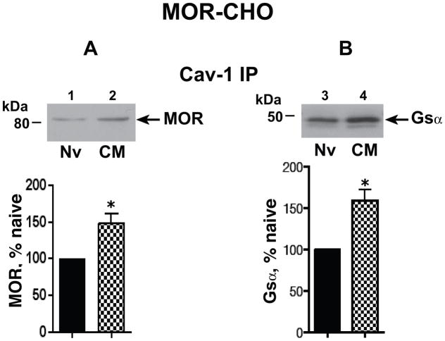
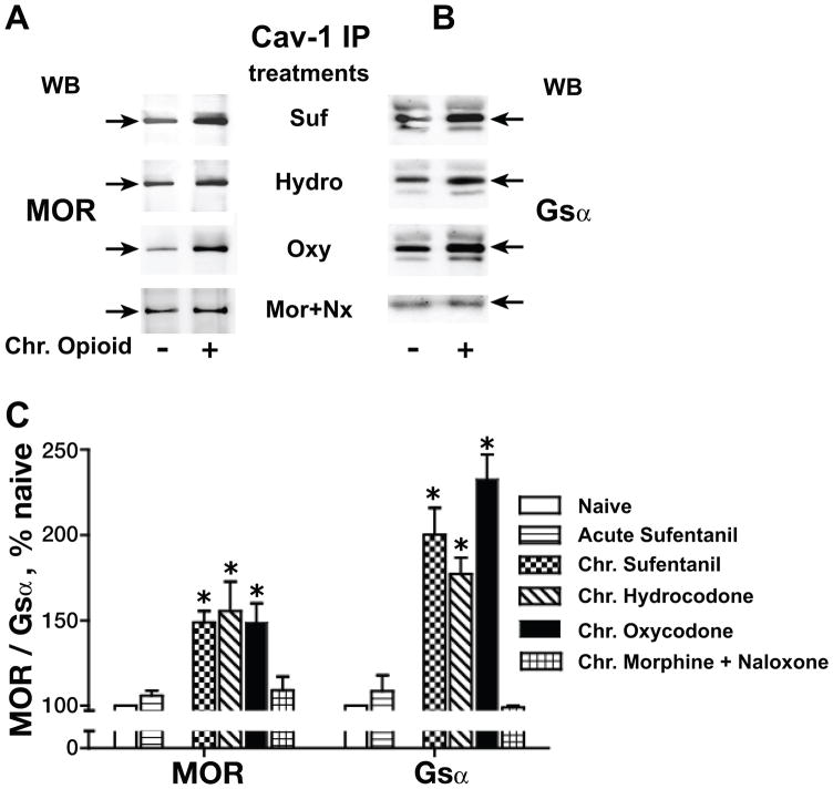
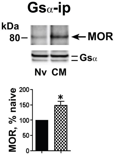
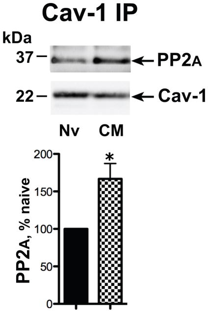
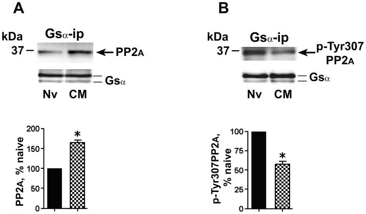
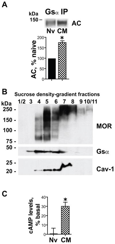
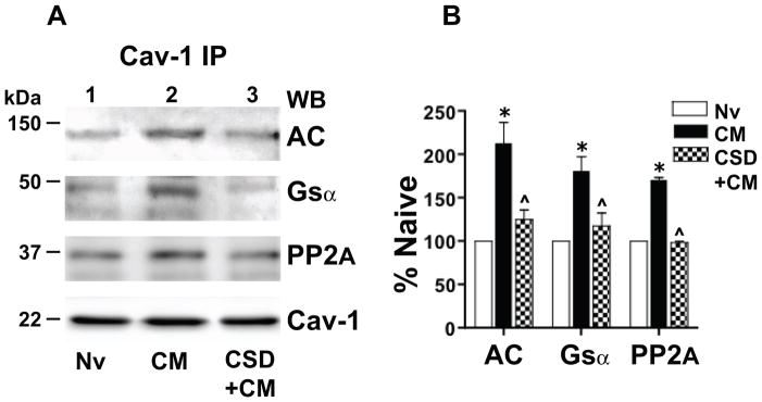
Caveolin-1 is the predominant structural protein of caveolae, a subset of (lipid) membrane rafts that compartmentalize cell signaling. Caveolin-1 binds most to G protein-coupled receptors and their signaling partners, thereby enhancing interactions among signaling cascade components and the relative activation of specific G protein-coupled pathways. This study reveals that chronic opioid exposure of μ-opioid receptor (MOR) expressing Chinese hamster ovary cells (MOR-CHO) and chronic in vivo morphine exposure of rat spinal cord augmented recruitment of multiple components of MOR-adenylyl cyclase (AC) stimulatory signaling by caveolin-1. Strikingly, in MOR-CHO and spinal cord, blocking the caveolin-1 scaffolding domain substantially attenuated the chronic morphine-induced increased interaction of caveolin-1 with MOR, Gsα, protein phosphatase 2A (PP2A), and AC. Chronic morphine treatment also increased interactions among the above signaling proteins, thus enabling sufentanil to stimulate (rather than inhibit) cAMP production within lipid membrane microdomains. The latter finding underscores the functionality of augmented interactions among MOR, Gs α, PP2A, and AC. In the aggregate, our data strongly suggest that augmented caveolin-1 scaffolding undergirds the ability of chronic opioids to recruit an ancillary signaling pathway by acting as an organizing template for MOR-Gs α-AC signaling and delimiting the membrane compartment(s) in which it occurs. Since caveolin-1 binds to a wide spectrum of signaling molecules, altered caveolin-1 scaffolding following chronic opioid treatment is likely to pertain to most, if not all, MOR signaling partners. The chronic morphine-induced trigger that augments caveolin-1 scaffolding could represent a seminal perturbation that initiates the wide spectrum of adaptations thought to contribute to opioid tolerance and dependence.

Keywords: caveolae; caveolin-1; opioid tolerance; opioids; μ-opioid receptor.


Conflict of interest statement

discloser The authors declare that they have no competing interests. Conflicts of interest: none

Figures

Figure 1
Figure 1
Chronic morphine increased the association of MOR and Gsα with Cav-1. Membranes were obtained from opioid naïve (Nv) and chronic morphine (CM) treated (1 μM, 48 h) MOR-CHO. The Triton insoluble fraction was isolated, solubilized, and subjected to immunoprecipitation with anti-Cav-1 antibody as described in methods. Following SDS PAGE and electrotransfer of proteins, regions of the nitrocellulose membrane corresponding to MOR, Gsα, and Cav-1 were cut out and Western blotted with their corresponding antibodies. Quantification of Western signals corresponding to A: MOR (≈80 kDa) and B, Gsα (≈45 kDa) in Cav-1 ip was normalized to their corresponding directly IPed Cav-1 protein. The content of MOR or Gsα in the Cav-1 IP obtained from chronic morphine-treated MOR-CHO is expressed as a percent of their content in Cav-1 IP obtained from opioid naïve cells. Following chronic morphine, the association of MOR and Gsα with Cav-1 within caveolae/Cav-1 scaffolds is significantly increased. n=4–5; *indicates p<0.05.
Fig. 2
Fig. 2
Effects of chronic morphine on Cav-1 scaffolding generalize to sufentanil (SUF), hydrocodone (HYDRO) and oxycodone (OXY). The Triton insoluble membrane fraction was obtained from opioid naïve, acute (5 min) sufentanil-treated and chronic sufentanil-, hydrocodone- or oxycodone-treated (1 μM, 48 h) MOR-CHO, after which it was solubilized and subjected to immunoprecipitation with anti-Cav-1 antibody. MOR (A) and Gsα (B) were visualized and quantified as described for Fig. 1. All co-IPs were normalized to their corresponding directly IPed Cav-1 protein. The content of MOR or Gsα in the Cav-1 IP from opioid treated MOR-CHO is expressed as a percent of their content in Cav-1 IP obtained from opioid naïve cells (C). n=3–5; *indicates p<0.05. The ability of chronic morphine to augment Cav-1 scaffolding of MOR and Gsα generalized to sufentanil, oxycodone, and hydrocodone. Chronic, not acute, opioid exposure is a prerequisite for the observed augmented Cav-1 scaffolding, which is eliminated by concomitant exposure to naloxone (Nx).
Figure 3
Figure 3
Chronic morphine augmented the association of MOR with Gsα. The Triton insoluble fraction obtained from membranes of opioid naïve (Nv) and chronic morphine (CM) treated MOR-CHO (1 μM, 48 h) was solubilized and subjected to immunoprecipitation using anti-Gsα antibodies. Co-IPed MOR was visualized by Western blotting. Quantification of band intensity was normalized to their corresponding directly IPed Gsα protein. The content of MOR in the Gsα IP obtained from chronic morphine-treated MOR-CHO is expressed as a percent of the MOR content of Gsα IP obtained from opioid naïve cells. Chronic morphine increased the association of MOR and Gsα within lipid raft compartments. n=4; *=p<0.01.
Figure 4
Figure 4
Chronic morphine augments the association of Cav-1 with PP2A. The Triton insoluble fraction obtained from membranes of opioid naïve (Nv) and chronic morphine (CM) treated MOR-CHO (1 μM, 48 h) was solubilized and subjected to immunoprecipitation using anti-Cav-1 antibodies. Co-IPed PP2A was visualized by Western blotting. Quantification of band intensity was normalized to their corresponding directly IPed Cav-1 protein. The content of PP2A in the Gsα IP obtained form chronic morphine-treated MOR-CHO is expressed as a percent of the Cav-1 content of Gsα IP obtained from opioid naïve cells. Chronic morphine increased association of PP2A and Cav-1 within caveolae/Cav-1 scaffolds. n=3; *=p<0.05.
Figure 5
Figure 5
Chronic morphine augments the association of activated (dephosphorylated) PP2A with Gsα. The Triton insoluble fraction of membranes of opioid naïve (Nv) and chronic morphine (CM) treated (1 μM, 48 h) MOR-CHO was solubilized and subjected to immunoprecipitation using anti-Gsα antibodies. A: Co-IPed PP2A was visualized by Western blotting using a pan anti-PP2A antibody. B: Gsα IP was obtained from an amount of Triton insoluble membrane protein identical to that used in panel A. The IP was Western blotted using an antibody specific for p-Tyr307PP2A. In both panels, quantification of band intensity was normalized by the amount of their corresponding directly IPed Gsα protein. The content of PP2A and p-Tyr307PP2A in the Gsα IP obtained from chronic morphine-treated MOR-CHO is expressed as a percent of their content in Gsα IP obtained from opioid naïve cells. Whereas the co-IP of PP2A with Gsα was augmented (≈65%) by chronic morphine, this treatment decreased by a similar magnitude (≈42%) the co-IP of p-Tyr307PP2A. In other words, the chronic morphine-induced increment in association of PP2A activity with Gsα is considerably greater than the increment in PP2A protein. n=5 for PP2A and n=3 for p-Tyr307PP2A; *=p<0.05.
Figure 6
Figure 6
Chronic morphine increases Gsα adenylyl cyclase (AC) association and augments MOR stimulation of cAMP formation in caveolae/Cav-1 scaffold fractions. A: The Triton insoluble fraction of membranes of opioid naïve (Nv) and chronic morphine (CM) treated MOR-CHO (1 μM, 48 h) was solubilized and subjected to immunoprecipitation using anti-Gsα antibodies. Co-IPed AC was visualized by Western blotting using an anti-AC common monoclonal antibody. Co-IPed AC was normalized to its corresponding directly IPed Gsα protein. The content of AC in the Gsα IP obtained from chronic morphine-treated MOR-CHO is expressed as a percent of its content in Gsα IP obtained from opioid naïve cells. Chronic morphine increased by ≈75% the magnitude of the co-IP of AC with Gsα within lipid raft compartments. n=3; *=p<0.05. B: Triton-insoluble components of MOR-CHO membranes were fractionated using sucrose density gradient centrifugation as described in Methods. The presence of MOR, Gsα and Cav-1 across the sucrose gradient was determined by Western blotting. The majority of Cav-1 was contained within the 20% and 30% sucrose gradients (corresponding to fractions 3–6). C: AC activity was quantified by measuring the synthesis of [32P]cAMP from [α-32P]ATP) in membrane raft fractions contained in the 20% and 30% sucrose gradients, corresponding to fractions 3–6 (fraction 3 was included since, in addition to Gsα, longer exposure of Westerns revealed the presence of MOR and Cav1, albeit at lower levels than the other included fractions obtained from opioid naïve and chronic morphine-treated MOR-CHO). Sufentanil (1 μM) stimulated [32P]cAMP formation in membrane raft fractions obtained from chronic morphine-treated (30±4%, *=p<0.002, n=5) but not opioid naïve MOR-CHO. Findings indicate that chronic morphine induced the emergence of MOR-coupled stimulatory AC signaling within membrane (lipid) rafts.
Figure 7
Figure 7
Chronic morphine augments association of Gsα, protein phosphatase 2A (PP2A) and adenylyl cyclase (AC) with Cav-1 in rat spinal cord. Their association is substantially attenuated/eliminated by blocking the Cav-1 scaffolding domain. A: The Triton insoluble fraction obtained was obtained from lumbar spinal cord membranes of opioid naïve rats (Nv, Lane 1), i.t. chronic morphine (CM)-treated rats (lane 2), or rats treated chronically with i.t. morphine concomitant with 10 μg of the Cav-1 scaffolding domain peptide CSD (CSD+CM) (lane 3; see Methods). The Triton insoluble fraction was solubilized and subjected immunoprecipitation using anti-Cav-1 antibodies. Following SDS PAGE and electrotransfer of proteins, regions of the nitrocellulose membrane corresponding to AC, Gsα, PP2A and Cav-1 were cut out and Western blotted with their corresponding antibodies. Quantification of band intensity was normalized to the amount of their corresponding directly IPed Cav-1 protein. B: The content of Gsα, PP2A and AC in the Cav-1 IP obtained from spinal cord of the above treated rats is expressed as a percent of their content in Cav-1 IP obtained from spinal cord of opioid naïve rats. A regimen of chronic in vivo morphine that produces spinal analgesic tolerance increased the association of Gsα, PP2A and AC with Cav-1 within caveolae/Cav-1 scaffolds of rat spinal cord. This increment was essentially eliminated by concomitant treatment with CSD. n=3; *=p<0.001 for CM vs. Nv; ^= p<0.01 for CM+CSD vs. CM.

References

    1. Allen JA, Yu JZ, Dave RH, Bhatnagar A, Roth BL, Rasenick MM. Caveolin-1 and lipid microdomains regulate Gs trafficking and attenuate Gs/adenylyl cyclase signaling. Mol Pharmacol. 2009;76:1082–1093. - PMC - PubMed
    1. Alvarez R, Daniels DV. A single column method for the assay of adenylate cyclase. Anal Biochem. 1990;187:98–103. - PubMed
    1. Bhatnagar A, Sheffler DJ, Kroeze WK, Compton-Toth B, Roth BL. Caveolin-1 interacts with 5-HT2A serotonin receptors and profoundly modulates the signaling of selected Galphaq-coupled protein receptors. J Biol Chem. 2004;279:34614–34623. - PubMed
    1. Boscher C, Nabi IR. Caveolin-1: role in cell signaling. Advances in experimental medicine and biology. 2012;729:29–50. - PubMed
    1. Bourova L, Kostrnova A, Hejnova L, Moravcova Z, Moon HE, Novotny J, Milligan G, Svoboda P. delta-Opioid receptors exhibit high efficiency when activating trimeric G proteins in membrane domains. J Neurochem. 2003;85:34–49. - PubMed

Publication types