. 2017 Feb;230(2):315-324.
doi: 10.1111/joa.12550. Epub 2016 Oct 11.

Reevaluation of the arterial blood supply of the auricle

Affiliations

Reevaluation of the arterial blood supply of the auricle

Isaac Zilinsky et al. J Anat. 2017 Feb.

Abstract

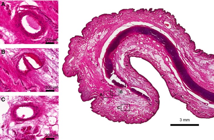
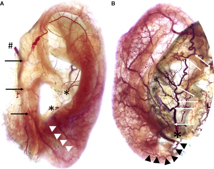
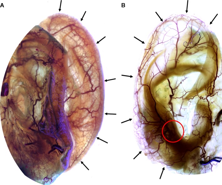
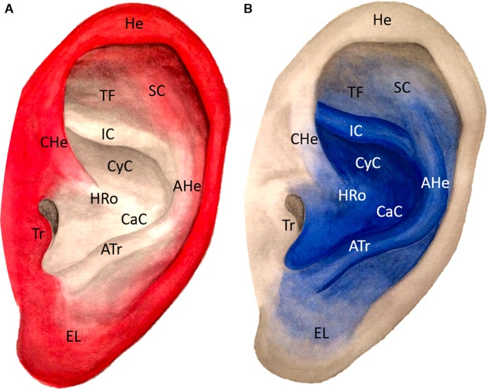
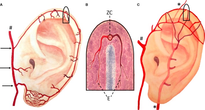
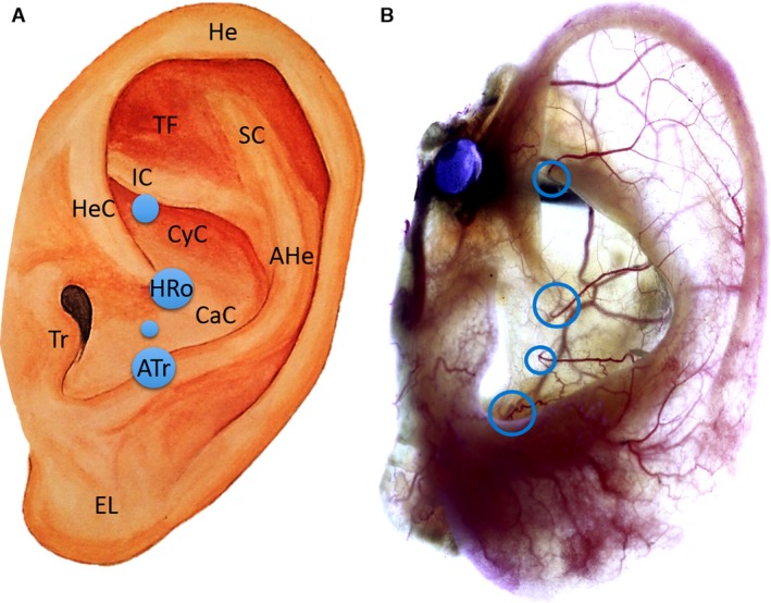
The anatomical basis for auricular flaps used in multiple aesthetic and reconstructive procedures is currently based on a random distribution of the underlying arterial network. However, recent findings reveal a systematic pattern as opposed to the present concepts. Therefore, we designed this study to assess the arterial vascular pattern of the auricle in order to provide reliable data about the vascular map required for surgical interventions. Sixteen human auricles from eight body donors (five females/three males, 84.33 ± 9.0 years) were investigated using the unique 'Spalteholz' method. After arterial injection of silicone, a complete transparency of the tissue was achieved and the auricular arteries and branches were visible. Qualitative and quantitative evaluation of the arterial vascular pattern was performed. The superior and the inferior anterior auricular artery provided the vascular supply to the helical rim, forming an arcade, i.e. helical rim arcade. On the superior third of the helical rim another arcade was confirmed between the superior anterior auricular artery and the posterior auricular artery (PAA), i.e. the helical arcade. The perforators of the PAA were identified lying in a vertical line 1 cm posterior to the tragus, supplying the concha, inferior crus, triangular fossa, antihelix and the earlobe. The results of this study confirmed the constant presence of the helical rim arcade (Zilinsky-Cotofana), consistent perforating branches of the PAA, and the helical arcade (Erdman), and will help and guide physicians performing auricular surgeries toward fast and simple procedures with optimal patient satisfaction.

Keywords: human auricle; posterior auricular artery; reconstruction of the auricle; superior temporal artery; vascular anatomy of the auricle; vascular supply.


Figures

Figure 1
Figure 1
Hematoxylin‐eosin (HE) staining of the helical rim taken from the middle third of the auricle. (A–C) Cross‐sections of arteries located on the helical rim, connecting the superior anterior auricular artery with the inferior anterior auricular artery forming the helical rim arcade (Zilinsky‐Cotofana arcade). Scale bar is presented in each picture.
Figure 2
Figure 2
Images show anterior (A) and posterior (B) view of left auricles stained according to ‘Spalteholz’ method. Image A: #Superficial temporal artery, black arrows mark superior/middle/inferior anterior auricular arteries, *Helical root and antitragal perforator, white arrowheads mark branch of antitragal perforator supplying the earlobe. Image B: *Posterior auricular artery, white arrows indicate the perforating and non‐perforating branches, black arrowheads mark the inferior anterior auricular artery running towards the earlobe.
Figure 3
Figure 3
Image showing the posterior (A) and the anterior (B) view of right auricles stained according to ‘Spalteholz’ method. Black arrows indicate the helical rim arcade (Zilinsky‐Cotofana arcade). Note the connection of the antitragal perforator with this arcade: red circle in image B.
Figure 4
Figure 4
Arterial vascular pattern of the superficial temporal artery is shown in red (Image A) and of the posterior auricular artery is shown in blue (Image B). AHe, Antihelix; Atr, Antitragus; CaC, Cavum conchae; CyC, Cymba conchae; EL, Earlobe; HRo, Helical root; He, Helix; HeC, Helical crus; IC, Inferior crus of antihelix; SC, Superior crus of antihelix; TF, Triangular fossa; Tr, Tragus.
Figure 5
Figure 5
Image A: Artistic drawing of the helical rim arcade (Zilinsky‐Cotofana arcade) and its position on the helical rim; #Superficial temporal artery, black arrows indicate superior/middle/inferior anterior auricular arteries. Image B shows a schematic drawing of a cross‐section of the helical rim in the superior third. Posterior branch is shown in dark red and anterior branch in bright red. Both branches form the transversally running helical arcade (E, Erdmann arcade). Anterior and posterior branches connect to the longitudinally running helical rim arcade (ZC, Zilinsky‐Cotofana arcade). Image C: Artistic drawing of the helical arcade (Erdmann arcade) and its position on the helical rim in the upper third of the helical rim; #Superficial temporal artery, *Posterior auricular artery.
Figure 6
Figure 6
Artistic drawing of left auricle (A) and left auricle stained according to ‘Spalteholz’ method (B). Image A: The blue circles indicate the location of the perforating branches of the posterior auricular artery, whereas the size of the blue circles represents their frequency observed in the investigated sample. Circle at the helical root (HRo) and at the antitragus (ATr) are 100%. AHe, Antihelix; Atr, Antitragus; CaC, Cavum conchae; CyC, Cymba conchae; EL, Earlobe; He, Helix; HeC, Helical crus; HRo, Helical root; IC, Inferior crus of antihelix; Tr, Tragus; SC, Superior crus of antihelix; TF, Triangular fossa. Image B: Blue circles show the anticrural, helical root, conchal and the antitragal perforators.
Figure 7
Figure 7
Image A: Tumor extirpation at the superior third of the helical rim of the right ear of a male patient. Image B: Ear during surgical reconstruction: ELBAF (Ear Lobe Based Advancement Flap) is raised based on blood supply from the earlobe. Image C: Ear after surgical reconstruction and wound closure. Image D: Ear 4 weeks after surgery.
Figure 8
Figure 8
Image A: Tumor extirpation at the middle third of the helical rim of the right ear of a male patient. Image B: Ear during surgical reconstruction using ELBAF (Ear Lobe Based Advancement Flap) technique with additional upper advancement of helical rim. Image C: Ear after surgical reconstruction and wound closure. Image D: Ear 4 weeks after surgery.

References

    1. Antia NH, Buch VI (1967) Chondrocutaneous advancement flap for the marginal defect of the ear. Plast Reconstr Surg 39, 472–477. - PubMed
    1. Converse JM (1958a) Reconstruction of the auricle. I. Plast Reconstr Surg Transplant Bull 22, 150–163. - PubMed
    1. Converse JM (1958b) Reconstruction of the auricle. II. Plast Reconstr Surg Transplant Bull 22, 230–249. - PubMed
    1. Crikelair GF (1956) A method of partial ear reconstruction for avulsion of the upper portion of the ear. Plast Reconstr Surg (1946) 17, 438–443. - PubMed
    1. Erdmann D, Bruno AD, Follmar KE, et al. (2009) The helical arcade: anatomic basis for survival in near‐total ear avulsion. J Craniofac Surg 20, 245–248. - PubMed