. 2016 Nov:13:90-98.
doi: 10.1016/j.ebiom.2016.10.001. Epub 2016 Oct 4.

Non-NAD-Like poly(ADP-Ribose) Polymerase-1 Inhibitors effectively Eliminate Cancer in vivo

Affiliations

Non-NAD-Like poly(ADP-Ribose) Polymerase-1 Inhibitors effectively Eliminate Cancer in vivo

Colin Thomas et al. EBioMedicine. 2016 Nov.

Abstract

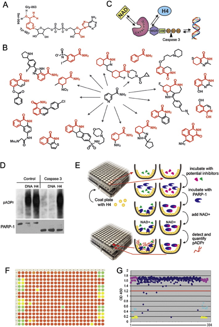
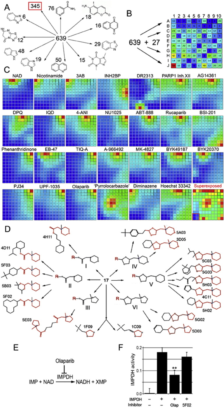
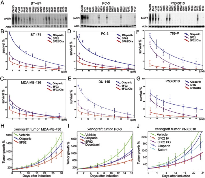
The clinical potential of PARP-1 inhibitors has been recognized >10years ago, prompting intensive research on their pharmacological application in several branches of medicine, particularly in oncology. However, natural or acquired resistance of tumors to known PARP-1 inhibitors poses a serious problem for their clinical implementation. Present study aims to reignite clinical interest to PARP-1 inhibitors by introducing a new method of identifying highly potent inhibitors and presenting the largest known collection of structurally diverse inhibitors. The majority of PARP-1 inhibitors known to date have been developed as NAD competitors. NAD is utilized by many enzymes other than PARP-1, resulting in a trade-off trap between their specificity and efficacy. To circumvent this problem, we have developed a new strategy to blindly screen a small molecule library for PARP-1 inhibitors by targeting a highly specific rout of its activation. Based on this screen, we present a collection of PARP-1 inhibitors and provide their structural classification. In addition to compounds that show structural similarity to NAD or known PARP-1 inhibitors, the screen identified structurally new non-NAD-like inhibitors that block PARP-1 activity in cancer cells with greater efficacy and potency than classical PARP-1 inhibitors currently used in clinic. These non-NAD-like PARP-1 inhibitors are effective against several types of human cancer xenografts, including kidney, prostate, and breast tumors in vivo. Our pre-clinical testing of these inhibitors using laboratory animals has established a strong foundation for advancing the new inhibitors to clinical trials.

Keywords: Cancer cells; Histone-dependent PARP-1 regulation; PARP-1; PARP-1 inhibitors; Poly(ADP-ribose).


Figures

Fig. 1
Fig. 1
Designing a new screening strategy to identify PARP-1 inhibitors. A. PARP-1 binds NAD + by NAD-binding pocket organized by three amino acids, including Gly-863, Ser-904 and Tyr-907, which mostly interact with the nicotinamide part of NAD. The parts of NAD used to develop PARP-1 inhibitors are shown in red. B. Most current PARP-1 inhibitors are developed from nicotinamide pharmacophore. C. Three ways of PARP-1 regulation: 1) competition with NAD for binding, 2) disruption of PARP-1 interaction with histones and 3) obstruction of binding with DNA. Arrowhead shows site of PARP-1 digestion by Caspase 3, which cleaves off DNA binding Zn-fingers of PARP-1, thus abolishing DNA-dependent PARP-1 activation. D. Interaction with the purified core histone H4 activates PARP1 in a DNA-independent manner. Full-length PARP-1 protein (left) and PARP1 protein cleaved by Caspase 3 (right) were preincubated with randomly broken DNA or core histone H4, followed by mixing with NAD. The product of PARP-1 enzymatic activity, poly(ADP-ribose), was detected after PAGE on a Western blot using anti-pADPr antibody. These data clearly demonstrate that the DNA-binding domain of PARP1 (Zn-fingers I and II) is not required for histone-dependent PARP1 activation. E. Schematic representation of the pipeline used to identify PARP-1 inhibitors. F-G. Data were visualized in a colour-coded table representing the 384-well plate in which potential inhibitors could be identified as green or yellow circles corresponding to wells that had minimal pADPr signal (F) or on a graph representing relative numerical value of this signal when compared to positive (yellow) or negative (purple) controls (G).
Fig. 2
Fig. 2
Identifying non-NAD-like small molecules inhibiting PARP-1 protein. A. Sorting out new PARP-1 inhibitors based on the presence of an obvious structural core, similar to known biologically active molecules. Eleven subgroups were identified. Structural cores and numbers of molecules falling in each group are indicated. B. Sorting out 639 new plus 27 known PARP-1 inhibitors and NAD based on 3D fingerprints, using the Canvas, ver. 1.6, program. Based on similarity of fingerprints, compounds were sorted to a 2D matrix containing 100 cells. The number of small molecules with all their 3D isomers in each cell is indicated. C. Comparison of molecules sorted in the matrix with each known PARP-1 inhibitor and NAD. Similarity is illustrated by heat map. Red corresponds to highest similarity and blue to absence of similarity. Name of inhibitor is indicated above the map. Bottom-right square represents a superimposure of 27 heat maps and reveals the area of matrix (labeled with red border) containing non-NAD-like compounds. D. Molecular structures of new PARP-1 inhibitors. Structural cores: I - 2-(N-methylmorpholino) acetate; II - 2-(N-methylpiperidin-1-yl)acetate; III - 2-(N-methylpyrrolidine-1-yl)acetate; IV - 1-((1,3-dioxolane-4-yl)methyl)N-methylmorpholino; V - 1-((1,3-dioxolane-4-yl)methyl) piperidine; VI - 1-((1,3-dioxolane-4-yl)methyl) N-methylpyrrolidine. E. Schematic illustration of IMPDH2 catalyzing reaction. F. Non-NAD-like inhibitors do not disrupt IMPDH2 activity. Graph showing IMPDH specific- activity in the IMPDH reaction with/without PARP-1 inhibitors. Column1: no recombinant human IMPDH2 added in the reaction. Column2: human IMPDH2 with 2 ul DMSO in the reaction. Column3: human IMPDH2 with 2 mM Olaparib in the reaction. Column 4: human IMPDH2 with 2 mM 5F02 in the reaction. **: P < 0.01.
Fig. 3
Fig. 3
PARP-1 inhibition and tumor cell suppression by classical NAD-mimetic and novel non-NAD-like PARP-1 inhibitors. (A) Non-NAD-like PARP-1 inhibitors block PARP-1 activity in human cells. A comparative analysis of PARP-1 activity in BT474, PC-3, and RCC cells cultured without and with classical PARP-1 inhibitors, 4ANI or PJ34, and new inhibitors identified in our screen. To detect pADPr on Western blot, we used mAb 10H antibody against pADPr. pAb antibody against Actin was used as a loading control. Reduction of pADPr was detected by Western blotting for inhibitor-treated cells relative to DMSO-treated cells. (B-G) New PARP-1 inhibitors suppress malignancy potential of cancer-derived cells: BT474 (breast cancer) (B); MDA-MB-436 (breast cancer) (C); PC-3 (prostate cancer) (D); DU145 (prostate cancer) (E); 789-P (RCC) (F); PNX0010 (RCC) (G). Calculation of cell survival rate in (B-G) was based on clonogenic cell survival assays. Cells were plated into 24-well plates. Cells were allowed to adhere overnight and were treated with a non-NAD-like inhibitor (5F02) (magenta), Olaparib (blue), and both (red) for 14 days. Colonies were counted and plotted on the graph. Data were fitted to exponential and logarithmic decay models using nonlinear curve fitting module of Statistica 7.0 software. The best fitting models for each inhibitor are represented on the chart. (H-J) Non-NAD-like PARP-1 inhibitors suppress tumor growth in vivo. 5F02 inhibitor suppresses growth of triple-negative breast cancer MDA-MB-436 (H), androgen-independent PC-3 (I), and renal cell carcinoma (PNX) (J) xenograft tumors in vivo. Ectopic MDA-MB-436 PC-3 or RCC xenograft tumors were established in 6-week-old male C·B17/Icr-scid mice (NOD.Cg-PrkdcscidIl2rgtm1Wjl/SzJ mice for MDA-MB-436). Animals were treated intraperitoneally with non-NAD-like inhibitor 5F02 (23 mg/kg), classical PARP-1 inhibitor Olaparib (Olap) (50 mg/kg), docetaxel (i.v. 12.5 mg/kg) for prostate xenografts, multi-targeted tyrosine kinase inhibitor (TKI) sunitinib (i.v. 40 mg/kg) for RCC xenograft tumors, or vehicle (PBS) 5 days a week. Values shown represent means (n = 5) + SEM. (J) 5F02 suppresses growth of RCC xenograft tumors. Xenograft tumors were established in 6-week-old male C·B17/Icr-scid mice using PNX0010 RCC cells generated from a clinical specimen of kidney cancer resistant to sunitinib treatment. Animals were treated with new PARP-1 inhibitor 5F02 (4 mg/kg intravenously (i.v.) or 6 mg/kg orally (p.o.)), classical PARP-1 inhibitor Olaparib (20 mg/kg, i.v.), sunitinib (40 mg/kg, p.o.), or vehicle 5 days a week. Values shown represent means (n = 5) + SEM. Data were fitted to exponential growth models using nonlinear curve fitting module of Statistica 7.0 software. (H-J) Data were fitted to exponential growth models using nonlinear curve fitting module of Statistica 7.0 software. Error bars correspond to standard deviation based on 5 repeats.

References

    1. Alberts B. Redefining cancer research. Science. 2009;325:1319. - PubMed
    1. Antolin A.A., Mestres J. Linking off-target kinase pharmacology to the differential cellular effects observed among PARP inhibitors. Oncotarget. 2014;5:3023–3028. - PMC - PubMed
    1. Antolín A.A., Jalencas X., Yélamos J., Mestres J. Identification of pim kinases as novel targets for PJ34 with confounding effects in PARP biology. ACS Chem. Biol. 2012;7:1962–1967. - PubMed
    1. Brenner J.C., Ateeq B., Li Y., Yocum A.K., Cao Q., Asangani I.A. Mechanistic rationale for inhibition of poly(ADP-ribose) polymerase in ETS gene fusion-positive prostate cancer. Cancer Cell. 2011;19:664–678. - PMC - PubMed
    1. Brown J.S., Kaye S.B., Yap T.A. PARP inhibitors: the race is on. Br. J. Cancer. 2016;114:713–715. - PMC - PubMed

MeSH terms