. 2016 Oct 11;11(10):e0164609.
doi: 10.1371/journal.pone.0164609. eCollection 2016.

Mitochondrial Superoxide Signaling Contributes to Norepinephrine-Mediated T-Lymphocyte Cytokine Profiles

Affiliations

Mitochondrial Superoxide Signaling Contributes to Norepinephrine-Mediated T-Lymphocyte Cytokine Profiles

Adam J Case et al. PLoS One. .

Abstract

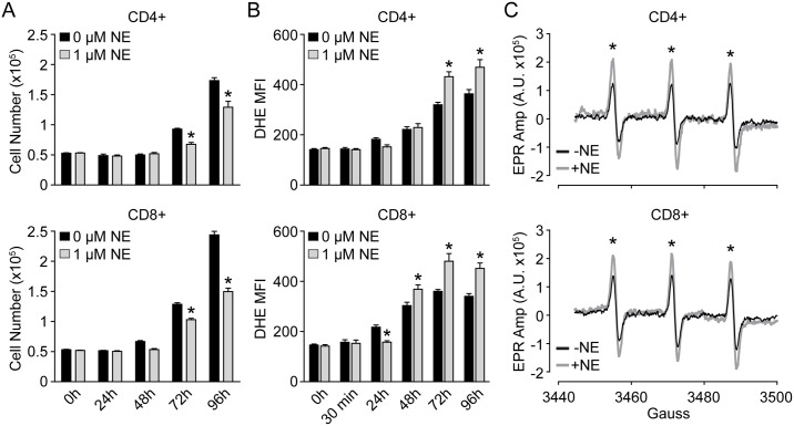
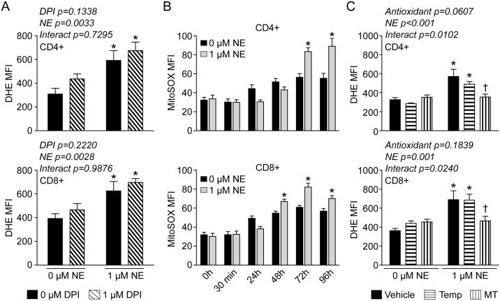
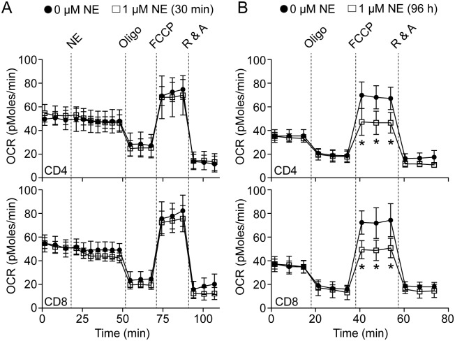
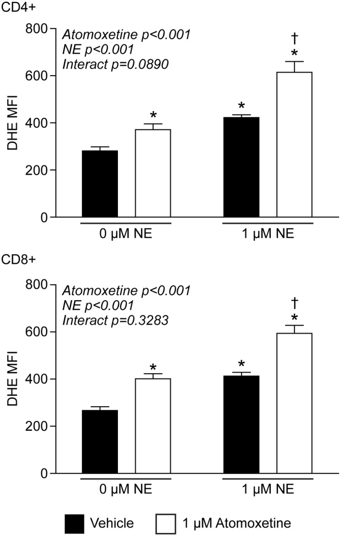
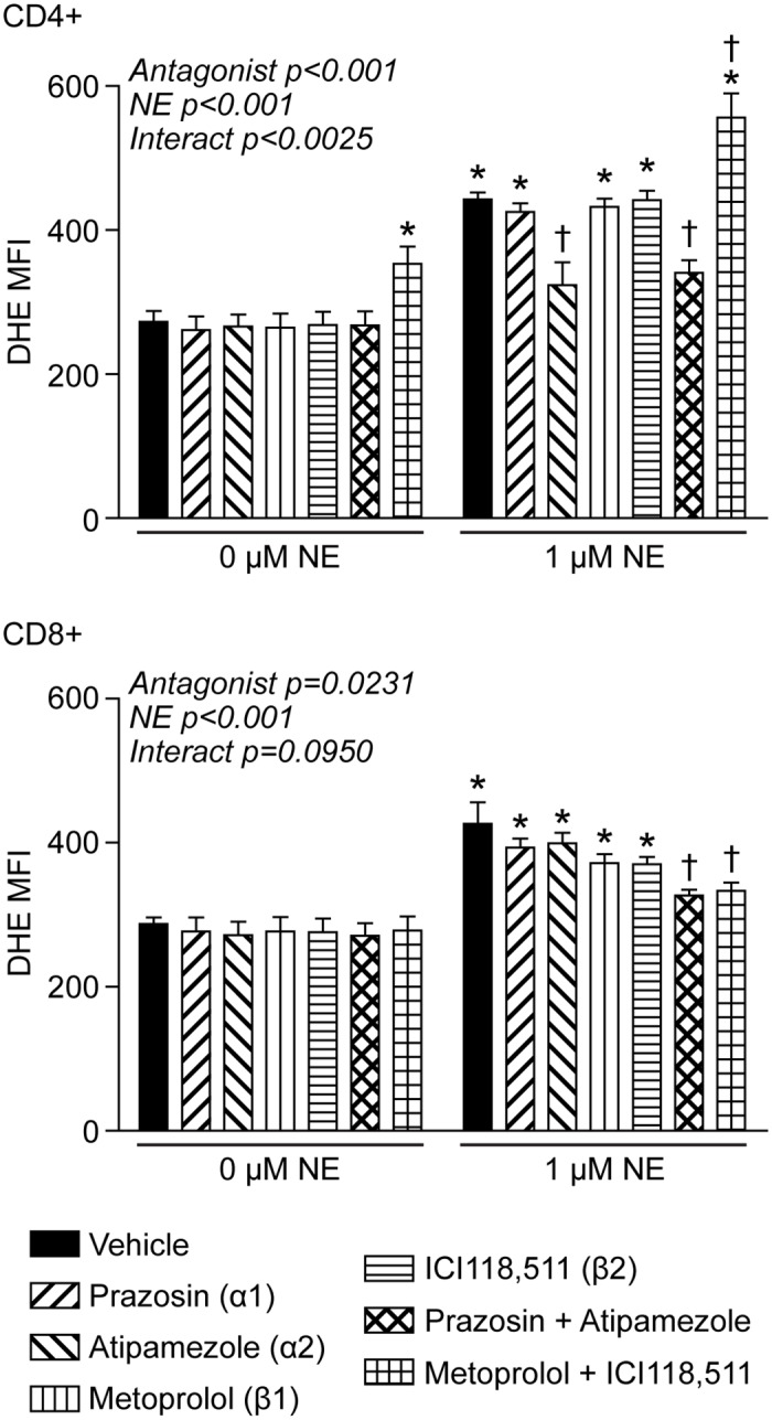
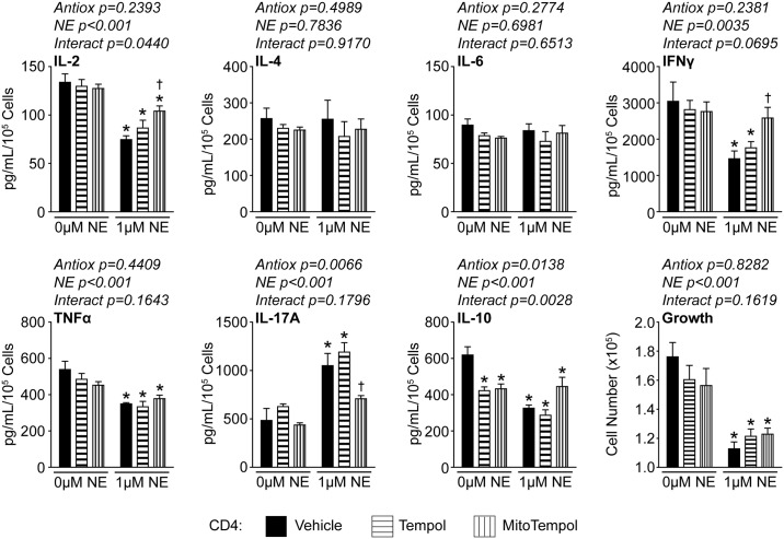
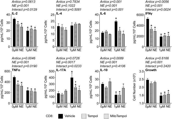
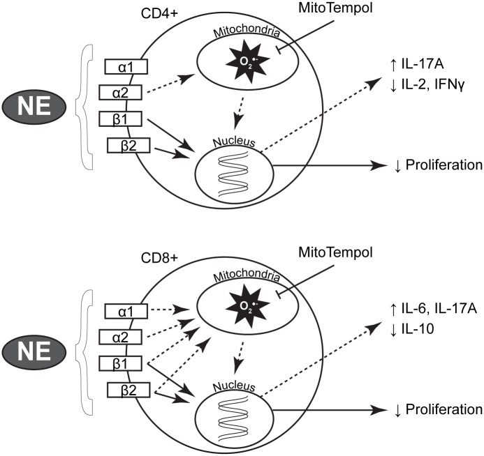
Norepinephrine (NE) produces multifaceted regulatory patterns in T-lymphocytes. Recently, we have shown that NE utilizes redox signaling as evidenced by increased superoxide (O2●-) causally linked to the observed changes in these cells; however, the source of this reactive oxygen species (ROS) remains elusive. Herein, we hypothesized that the source of increased O2●- in NE-stimulated T-lymphocytes is due to disruption of mitochondrial bioenergetics. To address this hypothesis, we utilized purified mouse splenic CD4+ and CD8+ T-lymphocytes stimulated with NE and assessed O2●- levels, mitochondrial metabolism, cellular proliferation, and cytokine profiles. We demonstrate that the increase in O2●- levels in response to NE is time-dependent and occurs at later points of T-lymphocyte activation. Moreover, the source of O2●- was indeed the mitochondria as evidenced by enhanced MitoSOX Red oxidation as well as abrogation of this signal by the addition of the mitochondrial-targeted O2●--scavenging antioxidant MitoTempol. NE-stimulated T-lymphocytes also demonstrated decreased mitochondrial respiratory capacity, which suggests disruption of mitochondrial metabolism and the potential source of increased mitochondrial O2●-. The effects of NE in regards to redox signaling appear to be adrenergic receptor-dependent as specific receptor antagonists could reverse the increase in O2●-; however, differential receptors regulating these processes were observed in CD4+ versus CD8+ T-lymphocytes. Finally, mitochondrial O2●- was shown to be mechanistic to the NE-mediated T-lymphocyte phenotype as supplementation of MitoTempol could reverse specific changes in cytokine expression observed with NE treatment. Overall, these studies indicate that mitochondrial metabolism and O2●--mediated redox signaling play a regulatory role in the T-lymphocyte response to NE.


Conflict of interest statement

The authors have declared that no competing interests exist.

Figures

Fig 1
Fig 1. NE provokes inverse effects on T-lymphocyte growth and O2●- levels.
T-lymphocytes were isolated, purified, and activated via CD3/CD28 stimulation in the presence of 1 μM NE. A. T-lymphocyte growth curves at various time points of ex vivo culture. N = 4. B. Quantification of DHE oxidation in CD4+ and CD8+ T-lymphocytes at various time points post-activation. N = 4. C. Representative EPR spectra showing amplitude (Amp) for T-lymphocytes stimulated in the presence or absence of 1 μM NE for 96 hours. N = 3. *p<0.05 vs. 0 μM NE by Student’s t-test at respective time points.
Fig 2
Fig 2. Mitochondrial O2●- is increased in T-lymphocytes treated with NE.
T-lymphocytes were isolated, purified, and activated via CD3/CD28 stimulation in the presence of 1 μM NE. A. Quantification of DHE oxidation in CD4+ and CD8+ T-lymphocytes 96 hours post-activation. Cells were incubated with DPI 1 hour prior to and during the incubation with DHE. N = 3. *p<0.05 vs. vehicle with 0 μM NE by 2-way ANOVA followed by Bonferroni post-hoc analysis. B. Quantification of MitoSOX red oxidation in CD4+ and CD8+ T-lymphocytes at various time points post-activation. N = 6. *p<0.05 vs. 0 μM NE by Student’s t-test at respective time points. C. Quantification of DHE oxidation in CD4+ and CD8+ T-lymphocytes at 96 hours post-activation. 1 μM Tempol (Temp) or MitoTempol (MT) were supplemented at time of plating and every 24 hours post-activation. N = 4. *p<0.05 vs. vehicle with 0 μM NE; p<0.05 vs. vehicle with 1 μM NE by 2-way ANOVA followed by Bonferroni post-hoc analysis.
Fig 3
Fig 3. T-lymphocyte mitochondrial reserve respiratory capacity is decreased with NE.
T-lymphocytes were isolated and purified. Cells were either immediately analyzed in a Seahorse Bioscience XFp extracellular flux analyzer with a 30-minute acute treatment of 1 μM NE, or activated via CD3/CD28 stimulation in the presence of 1 μM NE for 96 hours prior to analysis. A. Cumulative average data of oxygen consumption rate (OCR) measured over-time with acute treatment of NE. N = 9. B. Cumulative average data of OCR measured over-time in activated T-lymphocytes cultured in the presence of NE. N = 9. *p<0.05 vs. 0 μM NE by Student’s t-test at respective time points.
Fig 4
Fig 4. Inhibiting NE transport increases intracellular O2●- levels.
T-lymphocytes were isolated, purified, and activated via CD3/CD28 stimulation in the presence of NE and/or the norepinephrine transporter inhibitor atomoxetine. Quantification of DHE oxidation in CD4+ (upper) and CD8+ (lower) T-lymphocytes 96 hours post-activation. N = 4. *p<0.05 vs. vehicle with 0 μM NE; p<0.05 vs. vehicle with 1 μM NE by 2-way ANOVA followed by Bonferroni post-hoc analysis.
Fig 5
Fig 5. NE-mediated changes in O2●- are mediated via various adrenergic receptors.
T-lymphocytes were isolated, purified, and cells were activated via CD3/CD28 stimulation in the presence of 1 μM NE or the respective antagonist for 96 hours prior to analysis. Quantification of DHE oxidation in CD4+ (upper) and CD8+ (lower) T-lymphocytes activated in the presence of the treatments for 96 hours. N = 4. *p<0.05 vs. vehicle with 0 μM NE; p<0.05 vs. vehicle with 1 μM NE by 2-way ANOVA followed by Bonferroni post-hoc analysis.
Fig 6
Fig 6. Mitochondrial O2●- partially regulates CD4+ T-lymphocyte cytokine profiles.
CD4+ T-lymphocytes were isolated, purified, and activated via CD3/CD28 stimulation in the presence of NE, Tempol (1 μM), and/or MitoTempol (1 μM) for 96 hours. Media was harvested for cytokine analysis and results normalized to cell number. N = 5. *p<0.05 vs. vehicle with 0 μM NE; p<0.05 vs. vehicle with 1 μM NE by 2-way ANOVA followed by Bonferroni post-hoc analysis.
Fig 7
Fig 7. Mitochondrial O2●- partially regulates CD8+ T-lymphocyte cytokine profiles.
CD8+ T-lymphocytes were isolated, purified, and activated via CD3/CD28 stimulation in the presence of NE, Tempol (1 μM), and/or MitoTempol (1 μM) for 96 hours. Media was harvested for cytokine analysis and results normalized to cell number. N = 5. *p<0.05 vs. vehicle with 0 μM NE; p<0.05 vs. vehicle with 1 μM NE by 2-way ANOVA followed by Bonferroni post-hoc analysis.
Fig 8
Fig 8. Proposed NE signaling cascade in T-lymphocytes.
T-lymphocytes have been shown to express all adrenergic receptors, and NE has binding affinity to each isoform. In both CD4+ and CD8+ cells, NE appears to reduce proliferation via β-receptor canonical signaling (solid arrows) independent of redox events. CD4+ cells utilize the α2 receptor while CD8+ cells use all four adrenergic receptors to initiate redox signaling (dashed arrows) to affect specific subsets of cytokines. In both cells, the addition of MitoTempol can decrease this increase in mitochondrial O2●- and reverse the changes in specific cytokine expression.

References

    1. Guyenet PG. The sympathetic control of blood pressure. Nature reviews Neuroscience. 2006;7(5):335–46. Epub 2006/06/09. 10.1038/nrn1902 . - DOI - PubMed
    1. Zucker IH. Novel mechanisms of sympathetic regulation in chronic heart failure. Hypertension. 2006;48(6):1005–11. 10.1161/01.HYP.0000246614.47231.25 - DOI - PubMed
    1. Iyngkaran P, Anavekar N, Majoni W, Thomas MC. The role and management of sympathetic overactivity in cardiovascular and renal complications of diabetes. Diabetes & metabolism. 2013;39(4):290–8. Epub 2013/07/23. 10.1016/j.diabet.2013.05.002 . - DOI - PubMed
    1. Carter JR, Goldstein DS. Sympathoneural and adrenomedullary responses to mental stress. Comprehensive Physiology. 2015;5(1):119–46. Epub 2015/01/16. 10.1002/cphy.c140030 . - DOI - PMC - PubMed
    1. Nance DM, Sanders VM. Autonomic innervation and regulation of the immune system (1987–2007). Brain, behavior, and immunity. 2007;21(6):736–45. Epub 2007/05/01. 10.1016/j.bbi.2007.03.008 - DOI - PMC - PubMed

MeSH terms