. 2016 Dec:111:103-115.
doi: 10.1016/j.biomaterials.2016.09.029. Epub 2016 Sep 30.

Exosomes as biomimetic tools for stem cell differentiation: Applications in dental pulp tissue regeneration

Affiliations

Exosomes as biomimetic tools for stem cell differentiation: Applications in dental pulp tissue regeneration

Chun-Chieh Huang et al. Biomaterials. 2016 Dec.

Abstract

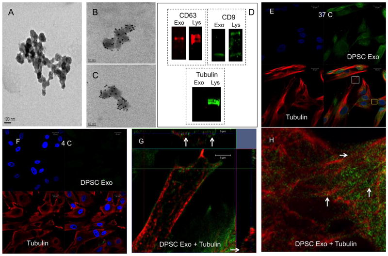
Achieving and maintaining safe and reliable lineage specific differentiation of stem cells is important for clinical translation of tissue engineering strategies. In an effort to circumvent the multitude of problems arising from the usage of growth factors and growth factor delivery systems, we have explored the use of exosomes as biomimetic tools to induce stem cell differentiation. Working on the hypothesis that cell-type specific exosomes can trigger lineage-specific differentiation of stem cells, we have evaluated the potential of exosomes derived from dental pulp cells cultured on under growth and odontogenic differentiation conditions to induce odontogenic differentiation of naïve human dental pulp stem cells (DPSCs) and human bone marrow derived stromal cells (HMSCs) in vitro and in vivo. Results indicate that the exosomes can bind to matrix proteins such as type I collagen and fibronectin enabling them to be tethered to biomaterials. The exosomes are endocytosed by both DPSCs and HMSCs in a dose-dependent and saturable manner via the caveolar endocytic mechanism and trigger the P38 mitogen activated protein kinase (MAPK) pathway. In addition, the exosomes also trigger the increased expression of genes required for odontogenic differentiation. When tested in vivo in a tooth root slice model with DPSCs, the exosomes triggered regeneration of dental pulp-like tissue. However, our results indicate that exosomes isolated under odontogenic conditions are better inducers of stem cell differentiation and tissue regeneration. Overall, our results highlight the potential exosomes as biomimetic tools to induce lineage specific differentiation of stem cells. Our results also show the importance of considering the source and state of exosome donor cells before a choice is made for therapeutic applications.

Keywords: Biomimetics; Dental pulp regeneration; Dental pulp stem cells; Exosomes; Regenerative endodontics.


Figures

Figure 1
Figure 1. Endocytosis of exosomes by DPSCs
(A) Representative TEM image of exosomes on nickel grids coated with carbon/fomvar film. This image also serves as secondary antibody negative control for the immune-gold labeled images in (B) and (C) that represent exosomes labeled for CD63 antigen with 10nm gold particles. (D) Immunoblots of protein extracts from exosome isolates and DPSCs showing the presence of marker proteins CD63 and CD9 and negative control tubulin (exo represents exosome and Lys represents cell lysate). (E) Representative confocal image of fluorescently labeled exosomes (green) endocytosed by DPSCs at 37°C counter stained with tubulin (red). (F) Representative confocal image of fluorescently labeled exosomes (green) endocytosed by DPSCs at 4°C counter stained with tubulin (red) (G) Orthogonal representation of z-stack confocal images of the area represented by white box in (E). Arrows point to exosomal presence on the microtubules in the x-z and y-z planes. (H) Higher magnification image of the area represented by the yellow box in (E) showing the presence of exosomes on the microtubules (white arrows).
Figure 2
Figure 2. Characterization of exosomal endocytic pathway
(A) Graph showing the dose dependent and saturable endocytosis of exosomes by DPSCs. Error bars represent standard deviation. The red line indicates a rectangular hyperbola fit to the data indicating that endocytosis follows a saturable binding curve suggesting a controlled mechanism. (B), (C) Representative confocal micrographs of untreated DPSCs (B) or DPSCs pre-treated with 2mM RGD peptide (C) and subjected to exosome endocytosis using fluorescently labeled exosomes (green) and counter stained with tubulin antibody (red). Note that the endocytosis was not blocked by the treatment. (D) Representative confocal micrograph of DPSCs containing endocytosed exosomes (green) stained for clathrin (red). No colocalization was observed between the two. (E) Representative confocal micrograph of DPSCs showing colocalization of endocytosed exosomes (green) with caveolin 1(red). (F) Orthogonal representation of z-stack confocal images showing three-dimensional colocalization of endocytosed exosomes with caveolin 1. Arrows point to regions of colocalization in the x-z and y-z planes. (G) Graph showing inhibition of exosomal endocytosis by MBCD at various concentrations. (H) Graph showing inhibition of exosomal endocytosis by DPSCs in the presence of heparin. For the graphs (G) and (H), (*) represents statistical significance with respect to the control group (P<0.05, student’s t-test)) and (#) represents statistical significance between the experimental groups (P<0.05, student’s t-test).
Figure 3
Figure 3. Endocytosis of exosomes triggers the activation of P38 MAPK pathway
(A) Representative confocal micrograph of DPSCs treated with control sample and immunostained with pP38 antibody (red). (B) Representative confocal micrograph of DPSCs treated with fluorescently labeled DPSC-Exo (green) immunostained with pP38 antibody (red). Note the enhanced nuclear translocation of pP38 (white arrows). (C) Representative confocal micrograph of DPSCs treated with fluorescently labeled DPSC-OD-Exo (green) immunostained with pP38 antibody (red). White arrows point to nuclear translocation of pP38. (D) Representative western blots showing the time dependent increase in pP38 levels. (E) Quantitation of western blot data for triplicate experiments showing normalized mean fold change in pP38 intensity. Total P38 expression was normalized to tubulin expression and the normalized P38 was used to obtain relative levels of pP38. Fold change was calculated for the exosome treatments with respect to the control. Data shows mean +/− SD. (*) Represents statistical significance (P<0.05, student’s t-test) with respect to control. (#) Represents statistical significance of DPSC-OD-Exo group with respect to DPSC-Exo group (P<0.05, student’s t-test). (F and G) Graph representing the expression levels of BMP2 and BMP9 respectively after 8 hours of treatment with DPSC-Exo in the absence and presence of P38 inhibitor SB203580 (SB). Note the reduction in the effect of exosome-mediated change in the presence of the inhibitor. Data shows mean +/− SD. (*) Represents statistical significance (P<0.05, student’s t-test) with respect to control. (#) Represents statistical significance between the exosome group and the exosome group with SB inhibitor (P<0.05, student’s t-test).
Figure 4
Figure 4. Binding of exosomes to ECM proteins
(A) Representative confocal micrograph of native DPSC generated ECM treated with control sample (no exosomes) and immunostained with fibronectin antibody (red). (B) Representative confocal micrograph of DPSC ECM treated with fluorescently labeled exosomes (green) immunostained with fibronectin antibody (red). Note the colocalization of the exosomes with fibronectin (white arrows). (C) Representative confocal micrograph of DPSC ECM treated with fluorescently labeled exosomes (green) that were pre-treated with 2mM RGD peptide to block integrins on the exosomal plasma membrane and immunostained with fibronectin antibody (red). Note the absence of green fluorescence indicating blocking of integrin mediated binding of exosomes to fibronectin. (D) Orthogonal representation of z-stack confocal images showing the co-localization of exosomes with fibronectin. Arrows point to colocalization in the x-z and y-z planes. (E) Graph representing the quantitation of exosomal binding to DPSC ECM and the effect of RGD peptide mediated integrin blocking on the binding. Note the significant decrease (*, P<0.05, student’s t-test) in the amount of bound exosome with RGD blocking, but not complete abrogation of binding. Data points represent mean of quadruplicate experiments +/− SD. (F) Graph representing the dose dependent and saturable binding of exosomes to type I collagen. Data points represent mean of quadruplicate experiments +/− SD. The red line represents a rectangular hyperbola fit to the data indicating saturable binding. (G, H, I) Representative transmission electron micrographs of exosomes alone (G), type I collagen alone (H) and exosome bound to type I collagen (I).
Figure 5
Figure 5. Histology and IHC of explant sections from the in vivo tooth root slice model
(A, A1, A2) Representative micrographs of explant sections from control (A), DPSC-Exo treated (A1) and DPSC-OD-Exo treated (C) immunostained with DMP1 antibody. Note the increased expression at the interphase between the soft tissue and the dentin (arrows) in exosome treated samples. Scale bar represents 50μm. (B, B1 and B2) Representative micrographs presented in the same order showing the localization of DPP. Only DPSC-OD-Exo treated samples showed a concentrated expression at the interface. Scale bar represents 50μm. (C, C1 and C2) Representative micrographs presented in the same order showing the localization of vWF. Only DPSC-OD-Exo treated samples showed increased expression. Scale bar represents 100μm. (D, D1, D2) Representative fluorescent micrographs showing autofluorescence in the red channel. Only DPSC-OD-Exo treated samples showed the presence of RBCs (white arrows) indicating the presence of capillaries within. Scale bar represents 10μm. (E, E1 and E2) Representative micrographs of H&E stained sections showing the overall morphology of the explants. Scale bar represents 20μm. (F, G) Representative micrographs showing absence of rabbit and mouse secondary antibody non-specific staining.
Figure 6
Figure 6. Fluorescence IHC of explant sections from the in vivo tooth root slice model
(A, A1, A2) Representative confocal micrographs of explant sections from control (A), DPSC-Exo treated (A1) and DPSC-OD-Exo treated (C) immunostained with BMP2 antibody. (B, B1, B2) Similarly presented representative confocal images showing the expression of TGFβ1. (C, C1, C2) Confocal images showing the expression of the transcription factor Runx2. (D, D1, D2) Confocal images showing the expression of the pro-angiogenic factor PDGF. Note the increased expression of all the proteins in the sections of samples treated with exosomes. Also note the increased expression in the DPSC-OD-Exo treated samples with respect to DPSC-Exo treated samples and the control. Scale bar represents 20μm in all images.
Figure 7
Figure 7. Endocytosis of DPSC exosomes by HMSCs
(A) Representative confocal micrograph of HMSCs treated with control sample and stained with phalloidin TRITC. (B) 3D representation of z-stack confocal images of HMSCs treated with fluorescently labeled DPSC exosomes (green) immunostained with tubulin antibody (red).

References

    1. Zara JN, Siu RK, Zhang X, Shen J, Ngo R, Lee M, et al. High doses of bone morphogenetic protein 2 induce structurally abnormal bone and inflammation in vivo. Tissue engineering Part A. 2011;17:1389–99. - PMC - PubMed
    1. Tannoury CA, An HS. Complications with the use of bone morphogenetic protein 2 (BMP-2) in spine surgery. The spine journal: official journal of the North American Spine Society. 2014;14:552–9. - PubMed
    1. Islam B, Khan SN, Khan AU. Dental caries: from infection to prevention. Medical science monitor: international medical journal of experimental and clinical research. 2007;13:RA196–203. - PubMed
    1. Cvek M. Prognosis of luxated non-vital maxillary incisors treated with calcium hydroxide and filled with gutta-percha. A retrospective clinical study. Endod Dent Traumatol. 1992;8:45–55. - PubMed
    1. Morito A, Kida Y, Suzuki K, Inoue K, Kuroda N, Gomi K, et al. Effects of basic fibroblast growth factor on the development of the stem cell properties of human dental pulp cells. Archives of histology and cytology. 2009;72:51–64. - PubMed

Publication types