

Acute epiploic appendagitis: Radiologic and clinical features of 12 patients
- PMID: 27728879
- PMCID: PMC5065630
- DOI: 10.1016/j.ijscr.2016.09.015
Acute epiploic appendagitis: Radiologic and clinical features of 12 patients
Abstract
Purpose: The aim of this work is to explain the clinical features and the imaging findings of primitive epiploic appendagitis in 12 patients.
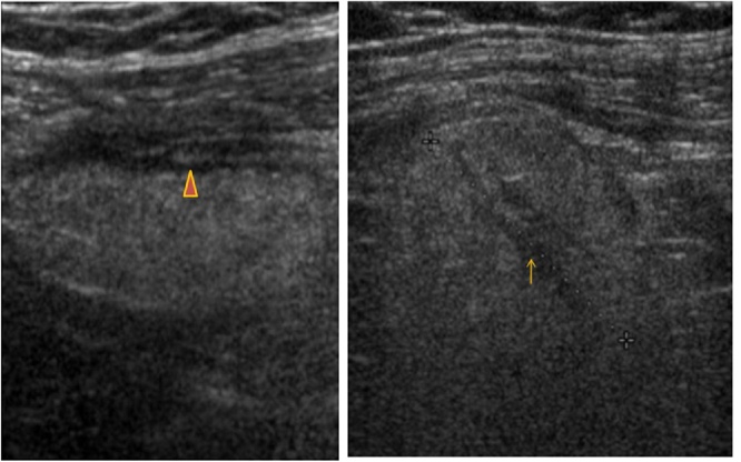
Materials and methods: Twelve patients were examined in 2 University hospitals between January 2011 and June 2016. Their medical charts have been reviewed. Nine patients have undergone enhanced CT examination and only two among them, have had at first an abdominal ultrasound.
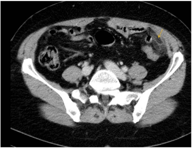
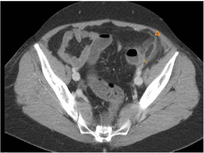
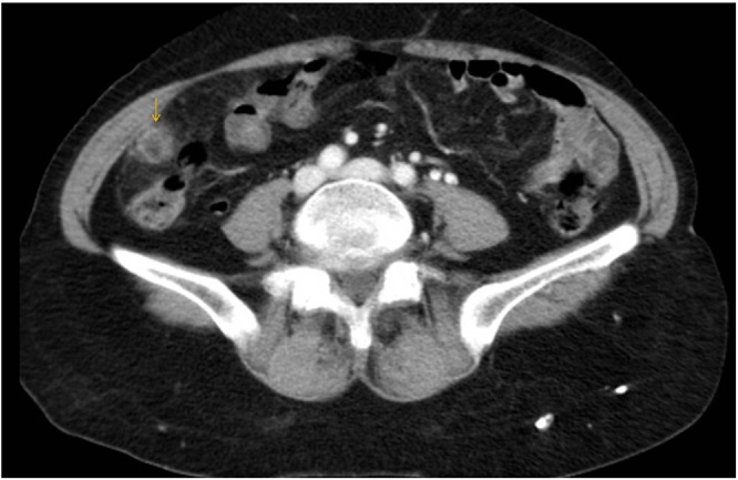
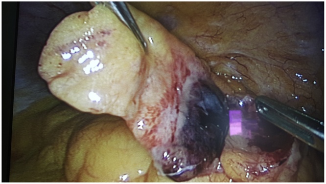
Results: The age ranged between 36 and 65 years old. All the patients consulted for an acute abdominal pain in most of the cases in the left iliac fossa with no elevated body temperature nor a significant elevation of the inflammation markers. Ultrasound features showed a hyper-echoic mass surrounded by a hypo-echoic peripheral ring. CT scan images showed a fat ovoid lesion that corresponds to the inflamed Appendix epiploica with a peripheral hyper-attenuating rim and in some cases the central "dot sign" referring to the thrombosed vessel. Only 4 patients underwent surgery.
Conclusion: For its non-specific clinical presentation, that can mimic other surgical affections, and its rather non-operative treatment, the diagnosis of epiploic appendagitis is crucial. Ultrasound and especially CT scan imaging are necessary for an accurate diagnosis.
Keywords: Acute abdomen; Computed tomography; Diagnosis; Management; Ultrasonography.
Copyright © 2016 The Author(s). Published by Elsevier Ltd.. All rights reserved.
Figures
References
-
- Saad J., Mustafa H.A., Elsani A.M., Alharbi F., Alghamdi S. Primary epiploic appendagitis: reconciling CT and clinical challenges. Indian J. Gastroenterol. 2014;33(5):420–426. - PubMed
-
- Singh A.K., Gervais D.A., Hahn P.F., Sagar P., Mueller P.R., Novelline R.A. Acute epiploic appendagitis and its mimics. Radiographics. 2005;25(6):1521–1534. - PubMed
-
- Singh A.K., Gervais D.A., Hahn P.F., Rhea J., Mueller P.R. CT appearance of acute appendagitis. Am. J. Roentgenol. 2004;183(5):1303–1307. - PubMed
LinkOut - more resources
Full Text Sources
Other Literature Sources
