Case Reports
. 2016 Oct 12;10(1):278.
doi: 10.1186/s13256-016-1061-x.

Symptomatic Cushing's syndrome and hyperandrogenemia in a steroid cell ovarian neoplasm: a case report

Affiliations
Case Reports

Symptomatic Cushing's syndrome and hyperandrogenemia in a steroid cell ovarian neoplasm: a case report

Ramy Sedhom et al. J Med Case Rep. .

Abstract

Background: Malignant steroid cell tumors of the ovary are rare and frequently associated with hormonal abnormalities. There are no guidelines on how to treat rapidly progressive Cushing's syndrome, a medical emergency.

Case presentation: A 67-year-old white woman presented to our hospital with rapidly developing signs and symptoms of Cushing's syndrome secondary to a steroid-secreting tumor. Her physical and biochemical manifestations of Cushing's syndrome progressed, and she was not amenable to undergoing conventional chemotherapy secondary to the debilitating effects of high cortisol. Her rapidly progressive Cushing's syndrome ultimately led to her death, despite aggressive medical management with spironolactone, ketoconazole, mitotane, and mifepristone.

Conclusions: We report an unusual and rare case of Cushing's syndrome secondary to a malignant steroid cell tumor of the ovary. The case is highlighted to discuss the complications of rapidly progressive Cushing's syndrome, an underreported and often unrecognized endocrine emergency, and the best available evidence for treatment.

Keywords: Cushing’s syndrome; Ectopic cortisol; Hyperandrogenemia; Steroid cell ovarian neoplasm.


Figures
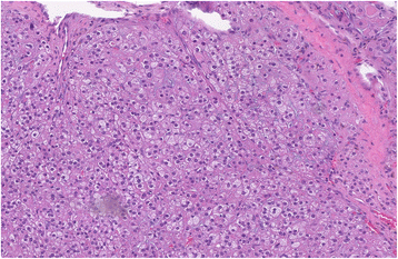
Fig. 1
Fig. 1
Hematoxylin and eosin stain with characteristic microscopic appearance and histology of steroid cell tumor not otherwise specified. Seen are large aggregates of polygonal to round tumor cells having distinct cell borders, central nuclei, and prominent nucleoli. Also present are delicate fibrous bands. Reinke crystals are absent
Fig. 2
Fig. 2
Higher-power view of Fig. 1. The cytoplasm of the tumor cells shows variably sized clear vacuoles, representing fat material
Fig. 3
Fig. 3
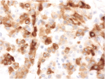
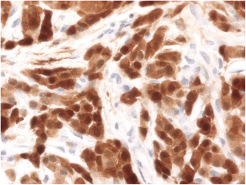
Immunohistochemical staining revealing inhibin positivity in the tumor cells, classifying the tumor as a sex cord-stromal tumor
Fig. 4
Fig. 4
Immunohistochemical staining revealing calretinin positivity in the tumor cells, revealing its steroid cell-secretory nature

References

    1. Young RH, Scully RE. Ovarian steroid cell tumors associated with Cushing’s syndrome: a report of three cases. Int J Gynecol Pathol. 1987;6(1):40–8. doi: 10.1097/00004347-198703000-00005. - DOI - PubMed
    1. Hayes MC, Scully RE. Ovarian steroid cell tumors (not otherwise specified): a clinicopathological analysis of 63 cases. Am J Surg Pathol. 1987;11(11):835–45. doi: 10.1097/00000478-198711000-00002. - DOI - PubMed
    1. Taylor HB, Norris HJ. Lipid cell tumors of the ovary. Cancer. 1967;20:1953–62. doi: 10.1002/1097-0142(196711)20:11<1953::AID-CNCR2820201123>3.0.CO;2-2. - DOI - PubMed
    1. Cope O, Raker JW. Cushing’s disease: the surgical experience in the care of 46 cases. N Engl J Med. 1955;253(4):119–27. doi: 10.1056/NEJM195507282530401. - DOI - PubMed
    1. Dunaif A, Scully RE. Case 22-1988—a 13-year-old girl with secondary amenorrhea, obesity, acanthosis nigricans, and hirsutism. N Engl J Med. 1988;318(22):1449–57. doi: 10.1056/NEJM198806023182207. - DOI - PubMed

Publication types

MeSH terms