Observational Study
. 2017 Apr 15;23(8):1998-2005.
doi: 10.1158/1078-0432.CCR-16-1371. Epub 2016 Oct 11.

Early Detection of Lung Cancer Using DNA Promoter Hypermethylation in Plasma and Sputum

Affiliations
Observational Study

Early Detection of Lung Cancer Using DNA Promoter Hypermethylation in Plasma and Sputum

Alicia Hulbert et al. Clin Cancer Res. .

Abstract

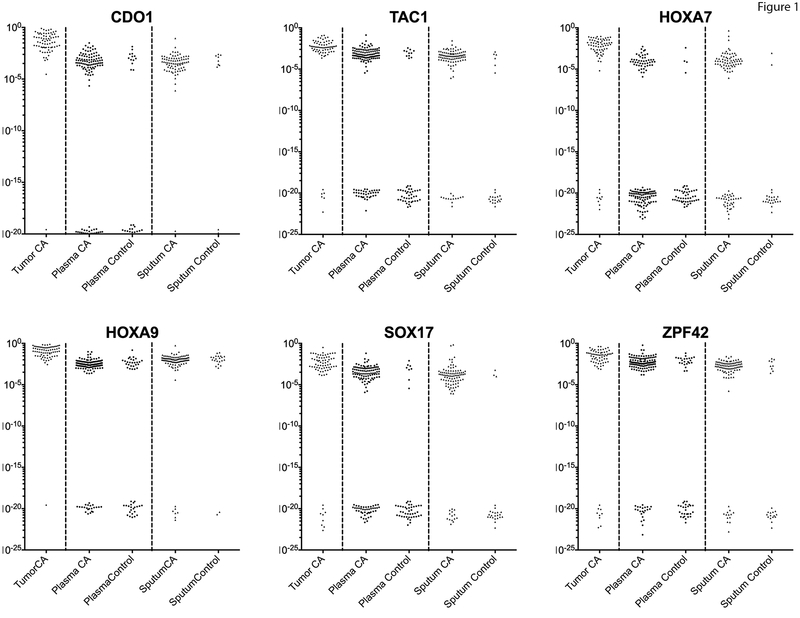
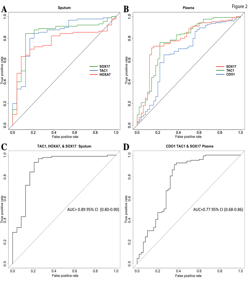
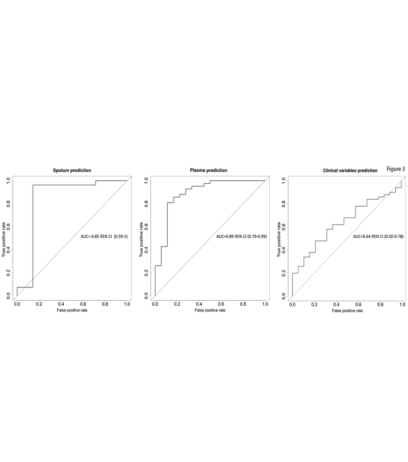
Purpose: CT screening can reduce death from lung cancer. We sought to improve the diagnostic accuracy of lung cancer screening using ultrasensitive methods and a lung cancer-specific gene panel to detect DNA methylation in sputum and plasma.Experimental Design: This is a case-control study of subjects with suspicious nodules on CT imaging. Plasma and sputum were obtained preoperatively. Cases (n = 150) had pathologic confirmation of node-negative (stages I and IIA) non-small cell lung cancer. Controls (n = 60) had non-cancer diagnoses. We detected promoter methylation using quantitative methylation-specific real-time PCR and methylation-on-beads for cancer-specific genes (SOX17, TAC1, HOXA7, CDO1, HOXA9, and ZFP42).Results: DNA methylation was detected in plasma and sputum more frequently in people with cancer compared with controls (P < 0.001) for five of six genes. The sensitivity and specificity for lung cancer diagnosis using the best individual genes was 63% to 86% and 75% to 92% in sputum, respectively, and 65% to 76% and 74% to 84% in plasma, respectively. A three-gene combination of the best individual genes has sensitivity and specificity of 98% and 71% using sputum and 93% and 62% using plasma. Area under the receiver operating curve for this panel was 0.89 [95% confidence interval (CI), 0.80-0.98] in sputum and 0.77 (95% CI, 0.68-0.86) in plasma. Independent blinded random forest prediction models combining gene methylation with clinical information correctly predicted lung cancer in 91% of subjects using sputum detection and 85% of subjects using plasma detection.Conclusions: High diagnostic accuracy for early-stage lung cancer can be obtained using methylated promoter detection in sputum or plasma. Clin Cancer Res; 23(8); 1998-2005. ©2016 AACR.


Conflict of interest statement

Conflict of Interest Statement: The authors of this manuscript report no relationship to disclose. The data in this manuscript were in part presented in the abstract/poster form at the 2015 Meeting of the American Association for Cancer Research (AACR) in Philadelphia, Pennsylvania 2015 Meeting of the International Association of Lung Cancer (IASLC) in Denver, Colorado

Figures

Figure 1.
Figure 1.. Methylation detection values of the studied genes.
This scatter plot shows the converted ΔCT methylation values in a logarithmic scale. These values show a bimodal distribution with the lower group the values corresponding to those samples with no detectable amplification (ND). The majority of lung tumor samples have high levels of methylation, as expected from the previous study. Plasma and sputum samples from cancer patients have detectable methylation which varies from levels nearing that of tumor samples to those at the limits of detection (10−5-10−6), while some patients are undetectable. The majority of controls have undetectable methylation at these loci, although some patients do have detectable methylation that is quantitatively similar to cancer patients. HOXA9 methylation is detectable in most control patients, especially in the sputum, suggesting this change is present in the lung epithelium and not as specific for the detection of cancer.
Figure 2.
Figure 2.. Receiver operator classification curves for lung cancer detection.
A. ROC curves comparing the 3 genes with the largest areas under the curve for sputum. B. ROC curves comparing the 3 genes with the largest areas under the curve for Plasma. C. ROC of the combined methylation status of the genes from sputum with the largest area under the curve. D. ROC of the combined methylation status of the genes from Plasma with the largest area under the curve. Abbreviations: area under the curve: AUC, 95 % confidence interval: 95% CI.
Figure 3.
Figure 3.. Receiver operator classification curves for cancer predictions.
ROC curves assessing the accuracy of the predictions for lung cancer performed on the testing subset by using as predictors the ΔCt values for all six genes, age, pack-year, COPD status and FVC values. The left plot is obtained using sputum samples, the middle one, using plasma samples and the right one, the ROC curve for the clinical predictors alone.

References

    1. National Lung Screening Trial Research Team, Aberle DR, Adams AM, Berg CD, Black WC, Clapp JD, et al. Reduced lung-cancer mortality with low-dose computed tomographic screening. N Engl J Med. 2011;365:395–409. - PMC - PubMed
    1. Tammemagi MC, Katki HA, Hocking WG, Church TR, Caporaso N, Kvale PA, et al. Selection criteria for lung-cancer screening. N Engl J Med. 2013;368:728–36. - PMC - PubMed
    1. Gierada DS, Pinsky P, Nath H, Chiles C, Duan F, Aberle DR. Projected outcomes using different nodule sizes to define a positive CT lung cancer screening examination. Journal of the National Cancer Institute. 2014;106. - PMC - PubMed
    1. Bach PB, Mirkin JN, Oliver TK, Azzoli CG, Berry DA, Brawley OW, et al. Benefits and harms of CT screening for lung cancer: a systematic review. JAMA. 2012;307:2418–29. - PMC - PubMed
    1. Esteller M, Sanchez-Cespedes M, Rosell R, Sidransky D, Baylin SB, Herman JG. Detection of aberrant promoter hypermethylation of tumor suppressor genes in serum DNA from non-small cell lung cancer patients. Cancer Res. 1999;59:67–70. - PubMed

Publication types

MeSH terms