. 2017 Jan 6;120(1):110-119.
doi: 10.1161/CIRCRESAHA.116.309977. Epub 2016 Oct 11.

SPEG (Striated Muscle Preferentially Expressed Protein Kinase) Is Essential for Cardiac Function by Regulating Junctional Membrane Complex Activity

Affiliations

SPEG (Striated Muscle Preferentially Expressed Protein Kinase) Is Essential for Cardiac Function by Regulating Junctional Membrane Complex Activity

Ann P Quick et al. Circ Res. .

Abstract

Rationale: Junctional membrane complexes (JMCs) in myocytes are critical microdomains, in which excitation-contraction coupling occurs. Structural and functional disruption of JMCs underlies contractile dysfunction in failing hearts. However, the role of newly identified JMC protein SPEG (striated muscle preferentially expressed protein kinase) remains unclear.

Objective: To determine the role of SPEG in healthy and failing adult hearts.

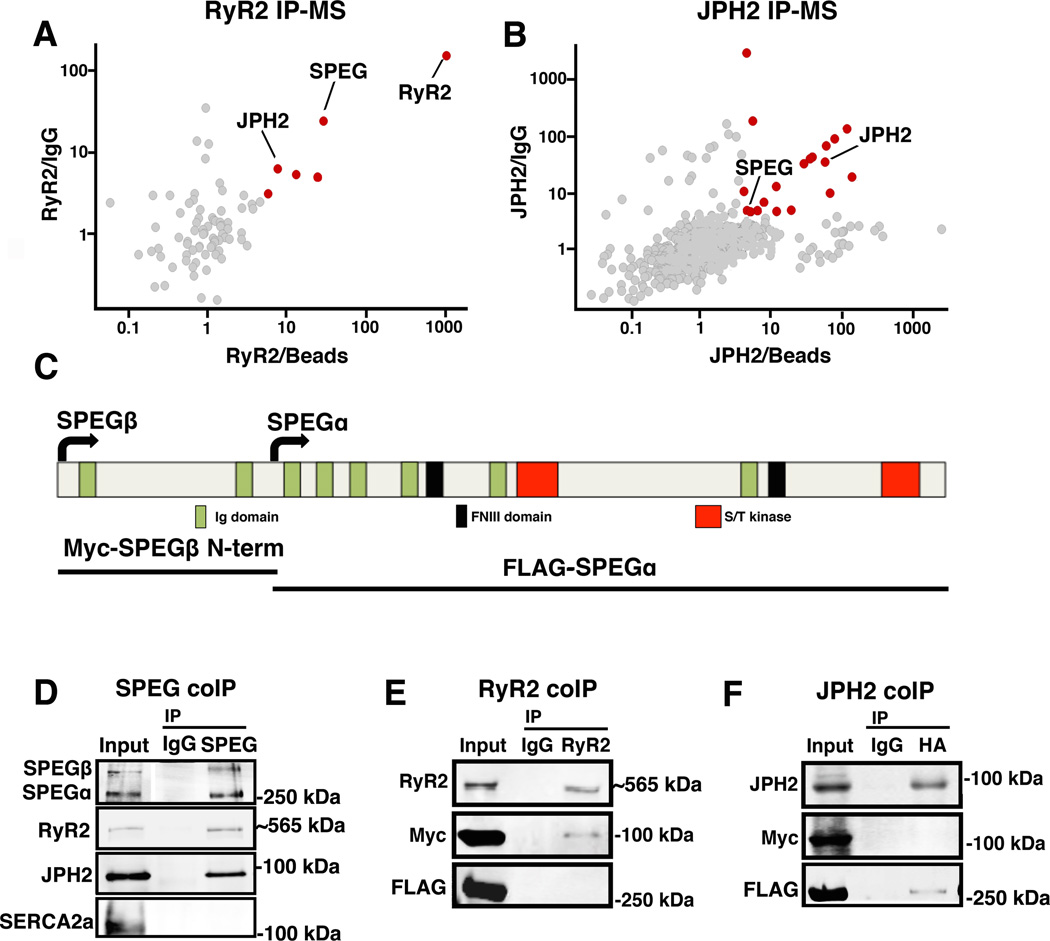
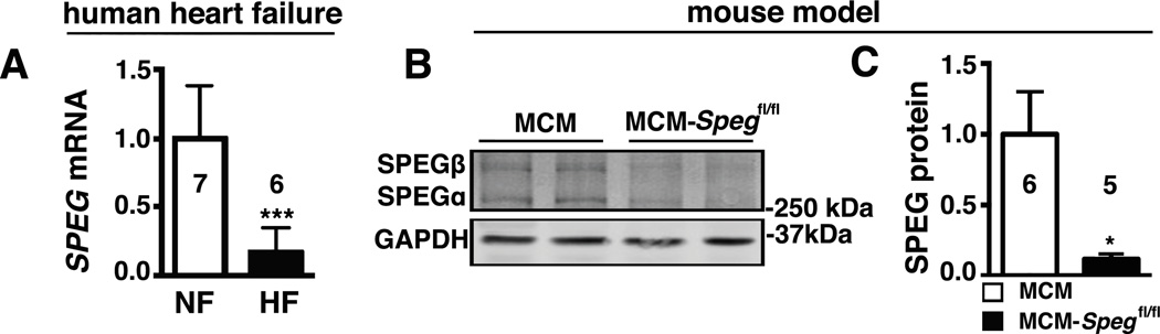
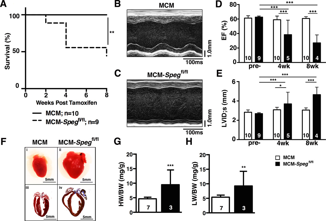
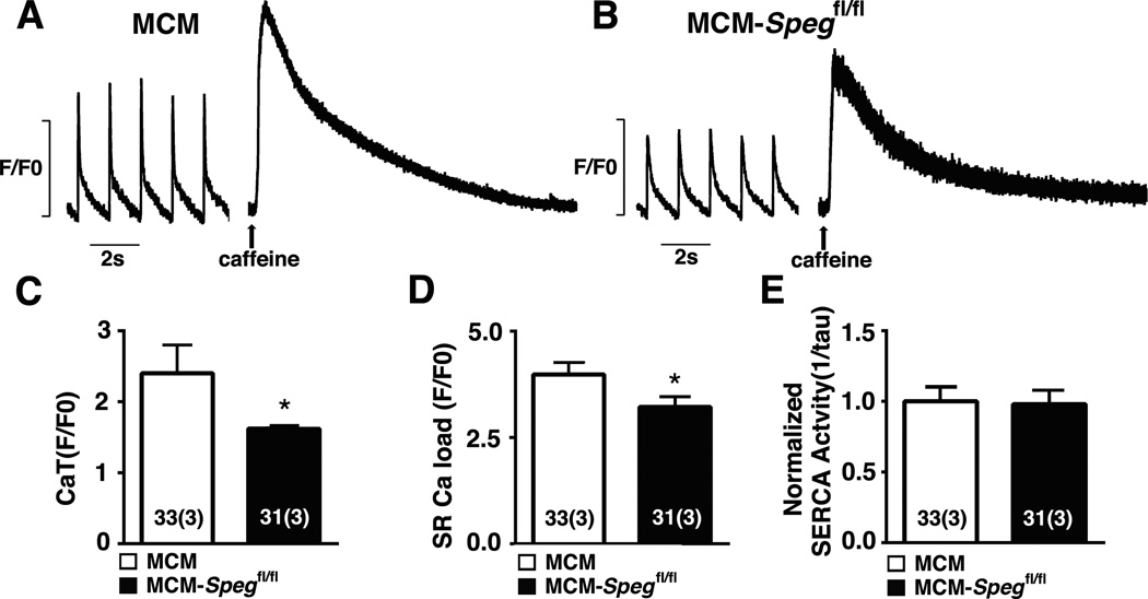
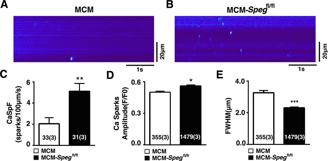
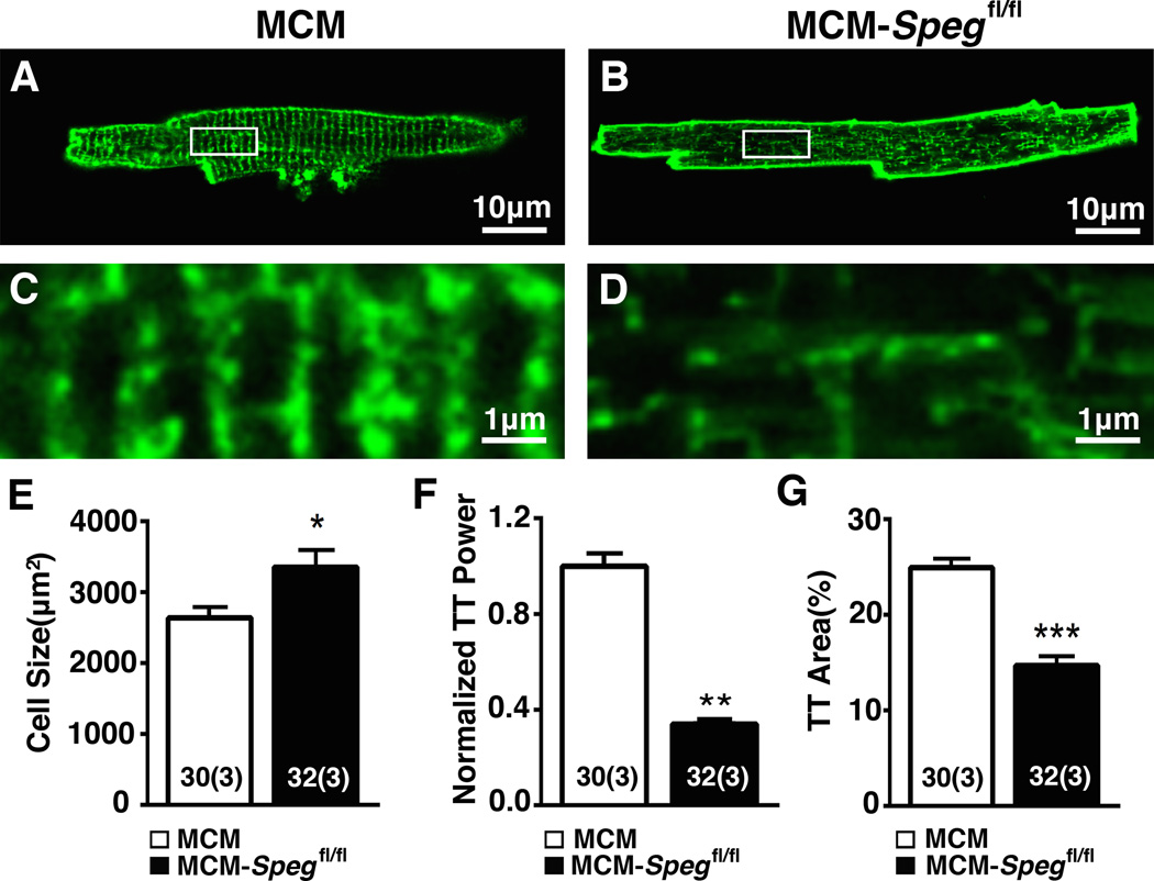
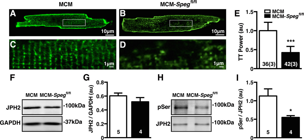
Methods and results: Proteomic analysis of immunoprecipitated JMC proteins ryanodine receptor type 2 and junctophilin-2 (JPH2) followed by mass spectrometry identified the serine-threonine kinase SPEG as the only novel binding partner for both proteins. Real-time polymerase chain reaction revealed the downregulation of SPEG mRNA levels in failing human hearts. A novel cardiac myocyte-specific Speg conditional knockout (MCM-Spegfl/fl) model revealed that adult-onset SPEG deficiency results in heart failure (HF). Calcium (Ca2+) and transverse-tubule imaging of ventricular myocytes from MCM-Spegfl/fl mice post HF revealed both increased sarcoplasmic reticulum Ca2+ spark frequency and disrupted JMC integrity. Additional studies revealed that transverse-tubule disruption precedes the development of HF development in MCM-Spegfl/fl mice. Although total JPH2 levels were unaltered, JPH2 phosphorylation levels were found to be reduced in MCM-Spegfl/fl mice, suggesting that loss of SPEG phosphorylation of JPH2 led to transverse-tubule disruption, a precursor of HF development in SPEG-deficient mice.

Conclusions: The novel JMC protein SPEG is downregulated in human failing hearts. Acute loss of SPEG in mouse hearts causes JPH2 dephosphorylation and transverse-tubule loss associated with downstream Ca2+ mishandling leading to HF. Our study suggests that SPEG could be a novel target for the treatment of HF.

Keywords: calcium signaling; heart failure; mass spectrometry; phosphorylation; proteomics.


Figures

Figure 1
Figure 1. SPEG directly binds to RyR2 and JPH2 within JMCs
(A–B) Unbiased proteomics analyses of putative RyR2 and JPH2 binding partners based on adult mouse heart co-immunoprecipitation of RyR2 and JPH2, respectively, coupled to mass spectrometry (IP-MS). Specific binding partners (red dots) were defined based on cut-off ratios of 3 and 4, respectively, of the MS intensity of each protein that came down with RyR2 or JPH2 IP vs. control IgG IP (y-axis) and RyR2 or JPH2 IP vs. beads only IP (x-axis). (C) Schematic overview of SPEG protein structure with key functional domains marked, and SPEG fragments used for binding-site analysis. (D) Western blots of reciprocal SPEG co-IP experiment from mouse heart showing that both RyR2 and JPH2 but not SERCA2a pulled down with SPEG. (E) RyR2, Myc-tagged SPEGβ N-terminal peptide, and FLAG-tagged SPEGα were co-expressed in HEK293 cells. Only SPEGβ N-terminal peptide was shown to bind the RyR2 pull-down. (F) JPH2 was co-expressed with Myc-tagged SPEGβ N-terminal peptide and FLAG-tagged SPEGα; only FLAG-tagged SPEGα was shown to bind the JPH2 pull-down.
Figure 2
Figure 2. SPEG is downregulated in human heart failure and a mouse model of cardiac myocyte specific inducible Speg knockout
(A) Quantitative real-time PCR analysis of SPEG mRNA transcript abundance in myocardium from patients suffering from HF or patients with normal heart function (NF). (B) SPEG western blots in hearts from inducible cardiac-specific SPEG-deficient mice (MCM-Spegfl/fl) and MCM-control mice, and (C) Quantification of SPEG levels in MCM and MCM-Spegfl/fl knockout mice. n, number of hearts, mice. * P<0.05, ***P<0.001.
Figure 3
Figure 3. SPEG downregulation causes heart failure
(A) Kaplan Meier survival plot showing percent survival of MCM and MCM-Spegfl/fl mice for 8 weeks after tamoxifen injection.(C–D) Representative M-mode echocardiography images from MCM and MCM-Spegfl/fl knockout mice at 8-weeks post-tamoxifen. Summary bar graphs showing (E) left ventricular ejection fraction (EF) and (F) left ventricular internal diameter in systole (LVID;s) at baseline (pre-) and 4 or 8 weeks after tamoxifen injection. (F) (i-ii) Representative images of whole hearts from MCM(i) and MCM-Spegfl/fl (ii) mice after tamoxifen and (iii-iv) Masson’s trichrome histological images of MCM(iii) and MCM-Spegfl/fl(iv) hearts. n, number of hearts/mice. * P<0.05, *** P<0.001 versus MCM control.
Figure 4
Figure 4. Abnormal systolic SR Ca2+ release in SPEG deficient cardiac myocytes
(A–B) Representative tracings of Ca2+ transient recordings after 1Hz pacing, subjected to caffeine at the end, in myocytes from MCM and MCM-Spegfl/fl knockout mice 8 weeks post tamoxifen. (C) Quantifications of Ca2+ transient amplitude, (D) sarcoplasmic reticulum (SR) Ca2+ load, and (E) SERCA2a activity. n, number of sparks (mice). * P<0.05.
Figure 5
Figure 5. Spontaneous RyR2-mediated Ca2+ release in cardiac myocytes with loss of SPEG
(A–B) Confocal microscopy line scans revealing increased number of Ca2+ sparks in MCM-Spegfl/fl mice 8 weeks post tamoxifen. (C) Bar graphs showing quantification of Ca2+ spark frequency (CaSpF), (D) Ca2+ spark amplitude, and (E) full-width of half max (FWHM). n, number of sparks (mice). * P<0.05, **P<0.01, ***P<0.001.
Figure 6
Figure 6. SPEG knockdown causes disrupted JMCs
(A–B) Ventricular myocytes isolated from MCM-Spegfl/fl mice or MCM-controls 8 weeks post tamoxifen stained with di-8-ANEPPS to visualize TT organization. (C–D) Enlarged image (corresponding to white box in panels A,B) showing disrupted TT structures in MCM-Spegfl/fl knockout mice. (E) Quantification of ventricular myocyte size, (F) normalized TT power, and (G) and TT area. n, number of sparks (mice). * P<0.05, **P<0.01, ***P<0.001.
Figure 7
Figure 7. Loss of TTs and JPH2 phosphorylation precedes heart failure in MCM-Spegfl/fl mice
(A–B) Representative images of ventricular myocytes isolated from MCM-Spegfl/fl mice or MCM controls 2 weeks post tamoxifen stained with di-8-ANEPPS and zoom (C–D) showing TT structure. (E) Quantification of TT Power. (F) Representative blot of total JPH2 with GAPDH as loading control from mouse heart lysates 2 weeks post tamoxifen and (G) corresponding quantification. (H) Representative image of JPH2 phosphoserine (pSer) Western blot normalized to JPH2 from immunoprecipitates from mice 2 weeks post tamoxifen and (I) quantification of phosphoserine normalized to total JPH2. Number in bars = number cells (mice); number hearts. * P<0.05.

Comment in

References

    1. Mozaffarian D, Benjamin EJ, Go AS, Arnett DK, Blaha MJ, Cushman M, Das SR, de Ferranti S, Despres JP, Fullerton HJ, Howard VJ, Huffman MD, Isasi CR, Jimenez MC, Judd SE, Kissela BM, Lichtman JH, Lisabeth LD, Liu S, Mackey RH, Magid DJ, McGuire DK, Mohler ER, 3rd, Moy CS, Muntner P, Mussolino ME, Nasir K, Neumar RW, Nichol G, Palaniappan L, Pandey DK, Reeves MJ, Rodriguez CJ, Rosamond W, Sorlie PD, Stein J, Towfighi A, Turan TN, Virani SS, Woo D, Yeh RW, Turner MB American Heart Association Statistics C, Stroke Statistics S. Executive summary: Heart disease and stroke statistics-2016 update: A report from the american heart association. Circulation. 2016;133:447–454. - PubMed
    1. Houser SR. Does protein kinase a-mediated phosphorylation of the cardiac ryanodine receptor play any role in adrenergic regulation of calcium handling in health and disease? Circ Res. 2010;106:1672–1674. - PubMed
    1. Ather S, Respress JL, Li N, Wehrens XH. Alterations in ryanodine receptors and related proteins in heart failure. Biochim Biophys Acta. 2013;1832:2425–2431. - PMC - PubMed
    1. van Oort RJ, Garbino A, Wang W, Dixit SS, Landstrom AP, Gaur N, De Almeida AC, Skapura DG, Rudy Y, Burns AR, Ackerman MJ, Wehrens XH. Disrupted junctional membrane complexes and hyperactive ryanodine receptors after acute junctophilin knockdown in mice. Circulation. 2011;123:979–988. - PMC - PubMed
    1. Reynolds JO, Chiang DY, Wang W, Beavers DL, Dixit SS, Skapura DG, Landstrom AP, Song LS, Ackerman MJ, Wehrens XH. Junctophilin-2 is necessary for t-tubule maturation during mouse heart development. Cardiovasc Res. 2013;100:44–53. - PMC - PubMed

MeSH terms