. 2016 Oct 25;113(43):12202-12207.
doi: 10.1073/pnas.1608679113. Epub 2016 Oct 11.

Retinol and ascorbate drive erasure of epigenetic memory and enhance reprogramming to naïve pluripotency by complementary mechanisms

Affiliations

Retinol and ascorbate drive erasure of epigenetic memory and enhance reprogramming to naïve pluripotency by complementary mechanisms

Timothy Alexander Hore et al. Proc Natl Acad Sci U S A. .

Abstract

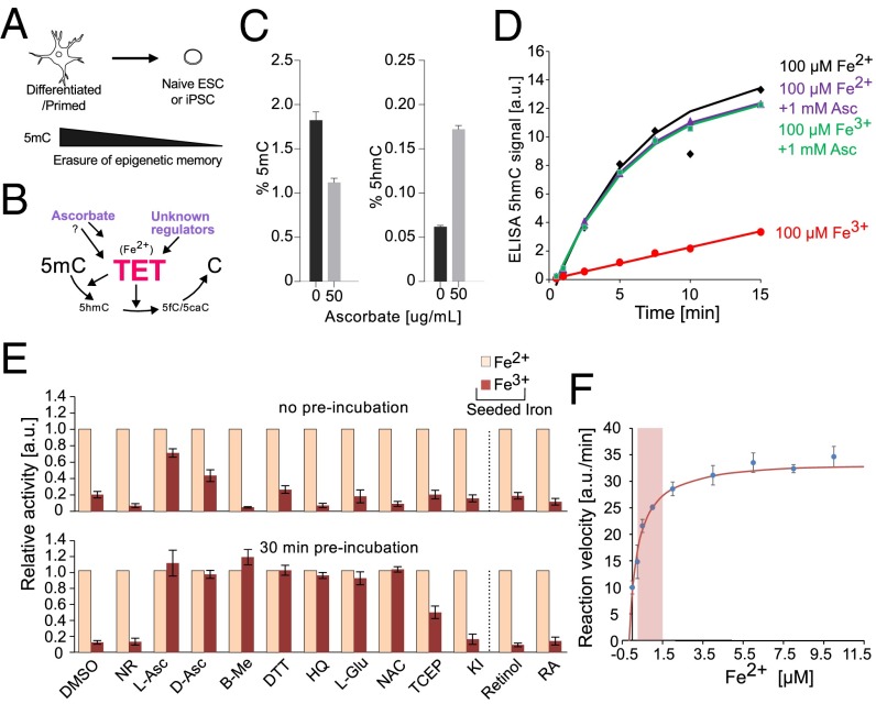
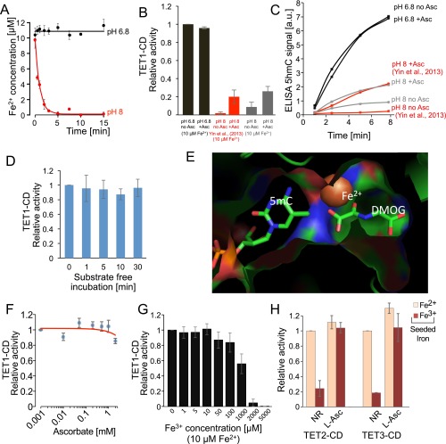
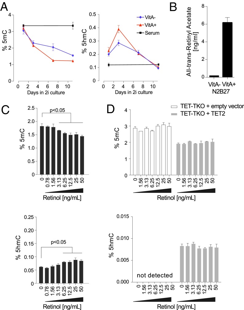
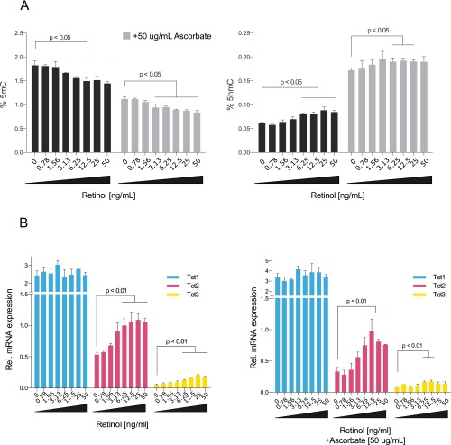
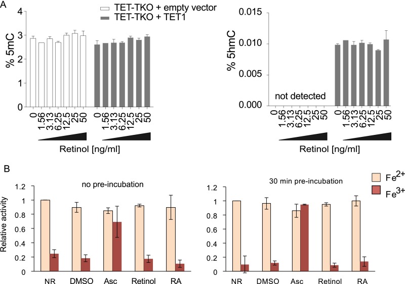
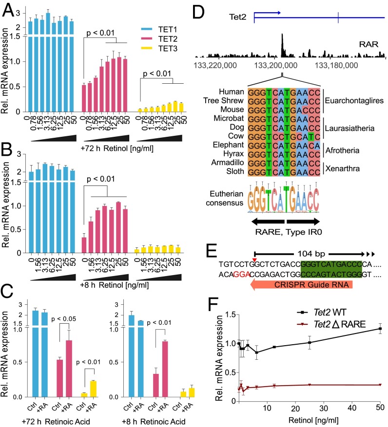
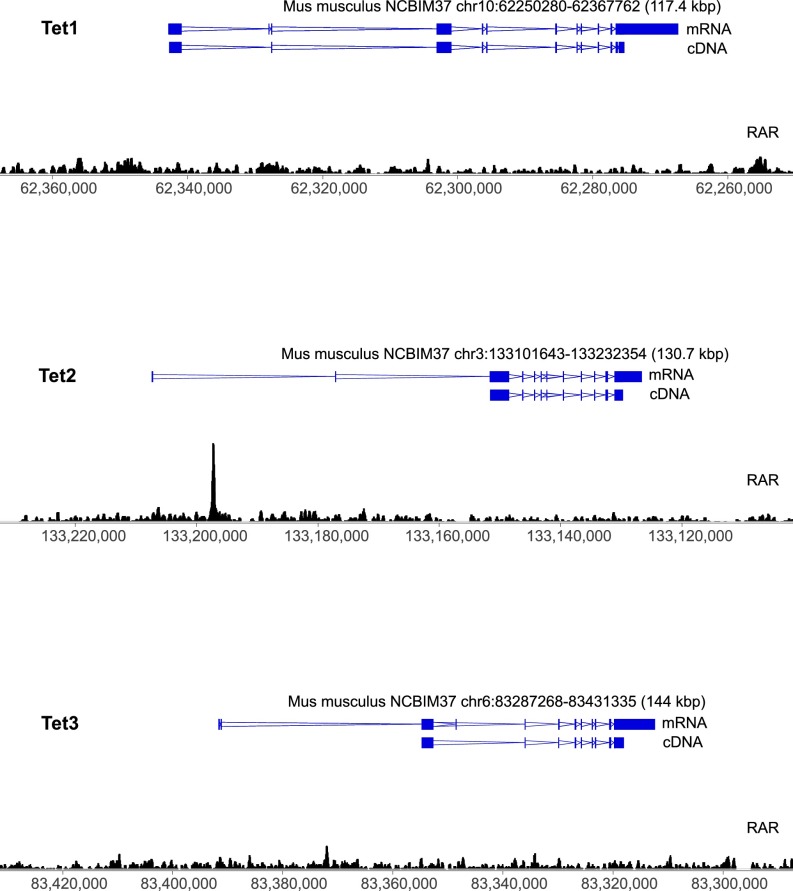
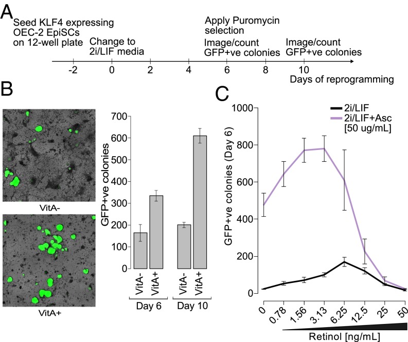
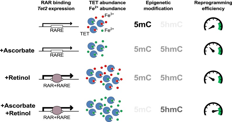
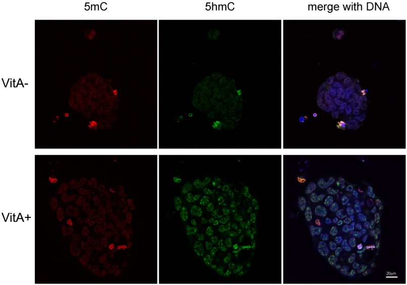
Epigenetic memory, in particular DNA methylation, is established during development in differentiating cells and must be erased to create naïve (induced) pluripotent stem cells. The ten-eleven translocation (TET) enzymes can catalyze the oxidation of 5-methylcytosine (5mC) to 5-hydroxymethylcytosine (5hmC) and further oxidized derivatives, thereby actively removing this memory. Nevertheless, the mechanism by which the TET enzymes are regulated, and the extent to which they can be manipulated, are poorly understood. Here we report that retinoic acid (RA) or retinol (vitamin A) and ascorbate (vitamin C) act as modulators of TET levels and activity. RA or retinol enhances 5hmC production in naïve embryonic stem cells by activation of TET2 and TET3 transcription, whereas ascorbate potentiates TET activity and 5hmC production through enhanced Fe2+ recycling, and not as a cofactor as reported previously. We find that both ascorbate and RA or retinol promote the derivation of induced pluripotent stem cells synergistically and enhance the erasure of epigenetic memory. This mechanistic insight has significance for the development of cell treatments for regenenerative medicine, and enhances our understanding of how intrinsic and extrinsic signals shape the epigenome.

Keywords: DNA methylation; TET; epigenetic memory; naive pluripotency; vitamin A/C.


Conflict of interest statement

W.R. and S.B. serve as consultants for Cambridge Epigenetix Ltd.

Figures

Fig. 1.
Fig. 1.
TET protein catalytic activity in vitro is rescued by the addition of Fe2+, ascorbate, or other antioxidants. (A) Major loss of DNA methylation, globally and at gene promoters, is associated with naïve pluripotent cells. (B) Demethylation during reprogramming is affected by the activity of the Fe2+-dependent TET hydroxylases, which create 5hmC and other oxidized derivatives (5fC and 5caC). However, what regulates TET levels is largely unknown, and the mechanism by which factors such as ascorbate affect TET-mediated oxidation are unclear. (C) Global levels of 5mC and 5hmC (%) in nESCs following 72 h of supplementation with 50 μg/mL ascorbate (± SD; n = 3). (D) Kinetics of TET1-CD–mediated 5hmC production when supplemented with 100 μM iron (Fe2+ or Fe3+) and 1 mM ascorbate (corresponds to 172.12 μg/mL). (E, Upper) Relative activity of TET1-CD when supplemented with 100 μM iron (Fe2+ or Fe3+) and various antioxidants. (E, Lower) The same experiment repeated, with the antioxidant and iron mix preincubated for 30 min before the addition of TET1-CD (± SD; n = 4). NR represents reaction conditions without added reducing agents. (F) Relative activity of TET1-CD at various Fe2+ concentrations encompassing those seen in cellular contexts (0.2–1.5 μM, indicated by a red box). The mean apparent dissociation constant (Kd) for this reaction was determined to be 0.41 ± 0.05 µM (n = 2).
Fig. S1.
Fig. S1.
Ascorbate rescues TET activity by Fe3+ reduction, not as a cofactor. (A) Fe2+ concentration within reaction mixes used to test TET activity. Although Fe2+ is stable for 15 min at pH 6.8 (black bar), it is rapidly lost at pH 8 (red bar) (± SD; n = 3). (B) Comparison of relative activity of TET1-CD (at 10 μM Fe2+) in the presence or absence of ascorbate at different reaction conditions. Black bars show enzyme activity (± SD; n = 3) under optimized conditions (at pH 6.8) as in Fig. 3, or using the conditions described by Yin et al. (23) at pH 8 (red) and our conditions, but at pH 8.0 (gray). (C) Reaction progression curves of 5hmC production from B. (D) Probing uncoupled partial reaction of the TET1-CD enzyme. TET1-CD was preincubated in the reaction mixture in the presence of oxoglutarate but the absence of substrate DNA. After the indicated time (on the x-axis), the reactions were started by the addition of DNA substrate, and the reaction kinetics were followed as before. Subsequently, the initial reaction rates were extracted and compared with the reaction rate without preincubation. (E) Catalytic center of human TET2 (PDB ID code 4NM6) (53) focused on the direct neighborhood of the bound catalytic iron. The Fe2+ ion is tightly contacted by oxoglutarate (a noncatalyzed analog, DMOG, is shown here) and 5mC, leaving no space for binding of an ascorbate molecule. (F) Relative activity of recombinant TET1-CD in the presence of increasing concentrations of ascorbate (± SD; n = 3). No stimulation of TET1 oxidation activity is seen; however, at high concentrations (2 mM ascorbate), weak suppression of TET activity is observed. (G) Competition between Fe2+ and Fe3+ for occupancy at the catalytic center. TET1-CD reactions containing 10 μM Fe2+ were supplemented with increasing amounts of Fe3+ ions. The initial rates of these reactions were compared with the oxidation reactions without the addition of Fe3+ (± SD; n = 4). (H) Relative activity of TET2-CD and TET3-CD when supplemented with 100 μM iron (Fe2+ or Fe3+) and either no reducing agent (NR) or l-ascorbate (l-Asc) (± SD; n = 3).
Fig. 2.
Fig. 2.
Retinol increases 5hmC and reduces 5mC in nESCs. (A) Global levels of 5mC and 5hmC (%) in serum-grown ESCs following 11 d of reprogramming in 2i/LIF medium either with vitamin A (VitA+) or without vitamin A (VitA−) (± SD; n = 3). (B) Retinyl acetate levels in VitA+/− 2i/LIF media formulations (± SD; n = 3). (C) Global levels of 5mC (Upper) and 5hmC (Lower) in nESCs supplemented with increasing levels of retinol for 72 h (black bars) (± SD; n = 3). (D) Global levels of 5mC (Upper) and 5hmC (Lower) in 2i/LIF-conditioned TET-TKO ESCs that were partially rescued by forced expression of TET2 (dark gray) and were exposed to retinol for 72 h (± SD; n = 3).
Fig. S2.
Fig. S2.
Effect of combined ascorbate and retinol treatment on nESCs. (A) Global levels of 5mC (Left) and 5hmC (Right) in nESCs supplemented with increasing levels of retinol for 72 h (black bars) (± SD; n = 3) and ascorbate plus increasing levels of retinol (gray bars). (B) Relative mRNA levels from TET1-3 in nESCs supplemented with retinol for 72 h (Left) and with retinol plus ascorbate (Right) (± SD; n = 3).
Fig. S3.
Fig. S3.
Retinol does not affect TET function posttranslation. (A) Global levels of 5mC (Left) and 5hmC (Right) in 2i/LIF-grown TET-TKO ESCs that were partially rescued by forced expression of TET1 (dark gray) and exposed to retinol for 72 h. Similar data were observed for TET2 (Fig. 2D). (± SD; n = 3). (B) Relative activity of TET1-CD when supplemented with 100 μM iron (Fe2+ or Fe3+) and retinol or RA, compared with ascorbate, and controls such as DMSO (carrier) or no reducing agent (NR). (± SD; n = 3).
Fig. 3.
Fig. 3.
Retinol enhances 5hmC in a TET2 and RA signaling-dependent manner. (A) Relative mRNA levels from TET1-3 in nESCs supplemented with retinol for 72 h (± SD; n = 3). (B) Relative mRNA levels from TET1-3 in nESCs supplemented with retinol for 8 h (± SD; n = 3). (C) Relative mRNA levels from TET1-3 in nESCs supplemented with RA (± SD; n = 3). (D) ChIP-seq of a pan-RAR antibody in ESCs (data analyzed from ref. 34). Underneath the major enrichment peak is an IR0-type RARE that is conserved throughout all mammalian superorders. (E) A 104-bp deletion was created encompassing the Tet2 RARE (green box) using a CRISPR guide RNA (orange arrow) downstream of a protospacer adjacent motif (red text). The full deletion coordinates are NCBI37, chr3:133197151–133197253. (F) Relative TET2 mRNA levels in retinol-supplemented nESCs with the Tet2 RARE deleted (Tet2 ΔRARE). Wild-type (WT) control cells are included for comparison (± SD; n = 3).
Fig. S4.
Fig. S4.
RAR binding at Tet1-3. ChIP-seq profiles using a pan-RAR antibody (black wiggle plot) in serum-grown ESCs following 8 h of treatment with RA (+8 h RA). Regions overlapping the Tet1, Tet2, and Tet3 genes are displayed. Exons are blue blocks and introns are double blue lines, with the apex of these lines representing the direction of transcription (data analyzed from ref. 34).
Fig. 4.
Fig. 4.
Retinol and ascorbate enhance reprogramming of epiblast stem cells to naïve pluripotency. (A) Experimental setup for EpiSC reprogramming experiments. (B) Oct4:GFP+ colonies following 6–10 d of reprogramming in VitA+/− 2i/LIF media. (Left) Representative microscopic field views at day 10 (4× magnification; the GFP channel is colored green and superimposed over a brightfield image of the same view). (Right) Frequency of reprogrammed colonies (± SD; n = 3). (C) Frequency of Oct4:GFP+ colonies after 6 d of reprogramming in VitA− 2i/LIF media with supplemented retinol and ascorbate (± SD; n = 3).
Fig. 5.
Fig. 5.
Retinol and ascorbate enhance DNA demethylation, 5hmC production, and pluripotent stem cell reprogramming by synergistic mechanisms. The RARE in the first intron of Tet2 allows increased expression of TET2 mRNA on stimulation of RA signaling (by retinol, retinyl acetate, or RA itself) and enhanced binding of the RAR (brown enzyme). In contrast, ascorbate increases the active iron (Fe2+, green circles) required for the TET catalytic center by reduction from Fe3+ (red circles). Together, retinol and ascorbate additively enhance 5hmC production, resulting in greater removal of methylation from DNA. The enhancing effect of ascobate and retinol on naïve pluripotent stem cell reprogramming is greater than the sum of their individual effects.
Fig. S5.
Fig. S5.
Retinol enhances 5hmC in human naïve ESCs. Immunofluorescence using antibodies specific to 5mC (red) and 5hmC (green) on human naïve ESCs grown with retinol (VitA+) and without retinol (VitA−).

References

    1. Ficz G, et al. FGF signaling inhibition in ESCs drives rapid genome-wide demethylation to the epigenetic ground state of pluripotency. Cell Stem Cell. 2013;13(3):351–359. - PMC - PubMed
    1. Habibi E, et al. Whole-genome bisulfite sequencing of two distinct interconvertible DNA methylomes of mouse embryonic stem cells. Cell Stem Cell. 2013;13(3):360–369. - PubMed
    1. Leitch HG, et al. Naive pluripotency is associated with global DNA hypomethylation. Nat Struct Mol Biol. 2013;20(3):311–316. - PMC - PubMed
    1. Yamaji M, et al. PRDM14 ensures naive pluripotency through dual regulation of signaling and epigenetic pathways in mouse embryonic stem cells. Cell Stem Cell. 2013;12(3):368–382. - PubMed
    1. Theunissen TW, et al. Systematic identification of culture conditions for induction and maintenance of naive human pluripotency. Cell Stem Cell. 2014;15(4):471–487. - PMC - PubMed

Publication types

MeSH terms

LinkOut - more resources