Review
. 2016 Oct 15;473(20):3401-3419.
doi: 10.1042/BCJ20160028.

Mechanism and disease association of E2-conjugating enzymes: lessons from UBE2T and UBE2L3

Affiliations
Review

Mechanism and disease association of E2-conjugating enzymes: lessons from UBE2T and UBE2L3

Arno F Alpi et al. Biochem J. .

Abstract

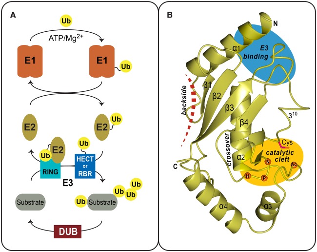
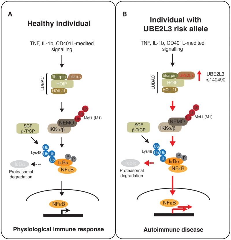
Ubiquitin signalling is a fundamental eukaryotic regulatory system, controlling diverse cellular functions. A cascade of E1, E2, and E3 enzymes is required for assembly of distinct signals, whereas an array of deubiquitinases and ubiquitin-binding modules edit, remove, and translate the signals. In the centre of this cascade sits the E2-conjugating enzyme, relaying activated ubiquitin from the E1 activating enzyme to the substrate, usually via an E3 ubiquitin ligase. Many disease states are associated with dysfunction of ubiquitin signalling, with the E3s being a particular focus. However, recent evidence demonstrates that mutations or impairment of the E2s can lead to severe disease states, including chromosome instability syndromes, cancer predisposition, and immunological disorders. Given their relevance to diseases, E2s may represent an important class of therapeutic targets. In the present study, we review the current understanding of the mechanism of this important family of enzymes, and the role of selected E2s in disease.

Keywords: E2-conjugating enzyme; Fanconi anaemia; UBE2L3; UBE2T; autoimmune disease; ubiquitin signalling.


Figures

Figure 1.
Figure 1.. Ubiquitin pathway and the E2 fold.
(A) Overview of the Ub pathway and the enzymes involved at each step: activation (E1), conjugation (E2), ligation (E3), and deubiquitination (DUB). The E1 mediates ubiquitin activation in an energy-consuming step. The ubiquitin thioester is then transferred onto a catalytic cysteine of the E2 enzyme. RING-type E3s form a non-covalent complex with the E2∼Ub thioester intermediate or, alternatively, ubiquitin is transferred to catalytic sites of HECT and RBR-type E3 ligases. The E3 enzymes ultimately catalyze ubiquitination of a substrate lysine. Ubiquitin signals can also be extended to form polyubiquitin chains. Finally, DUBs catalyze the removal of ubiquitin. (B) Ribbon diagram of UBE2D2 (PDB 2ESK) as a representative of the UBC fold conserved among ubiquitin E2s. Secondary structure elements and the termini of the domain are indicated. Also depicted is the location of the active-site cysteine within the catalytic cleft (yellow oval), the E3-binding region (blue oval), the cross-over helix that stabilizes ‘closed’ conformers of the E2∼Ub thioester intermediate and location of the E2 backside-binding surface (red dashed line).
Figure 2.
Figure 2.. The Fanconi anaemia pathway.
The FA pathway comprising 20 proteins is grouped into two classes: class I defines an upstream ubiquitin signalling module, and class II consists of downstream DNA repair proteins. The FA core complex is activated by stalled replication forks and co-operates with the E2-conjugating enzyme UBE2T (T) to monoubiquitinate the heterodimer FANCI-FANCD2 (I-D2). The RING E3 ligase FANCL (L) is the catalytic subunit, with associated FANCB (B) and FAAP-100 (F-100). Recruitment to stalled forks is mediated by FANCM (M), FAAP-10, -16, and -24. Monoubiquitinated I–D2 then promotes DNA repair by coordinating TLS (translesion synthesis) polymerase, nucleases, and HR-dependent pathways.
Figure 3.
Figure 3.. Schematic overview of identified UBE2T gene alterations in FA patients.
(A) Genomic locus of UBE2T with intron/exon boundaries. Start codon in exon 2, stop codon in exon 7, and AluY repeats (red boxes) are indicated. UBE2T encodes a 197 aa protein with catalytic Cys86 in the UBC fold (dark blue). (B) Maternal missense mutation identified in patient PNGS-252 resulting in Gln2 to Glu2 amino acid substitution. (C) The c.180+5G>A splice donor site, identified in patient PNGS-255, initiating a frame shift and premature stop codon resulting in a truncated UBE2T protein. Genome alterations identified patient 100166/1 showing the paternal AluY-mediated deletion (D) and maternal AluY-mediated duplication (E). The maternal allele encodes an mRNA for a shorter UBE2T protein with a functional UBC fold.
Figure 4.
Figure 4.. UBE2T amplifications described in human cancer studies.
Graphic summary of frequencies of UBE2T alterations in cancers, mainly amplifications based on expression profile data from a panel of cancer cell lines deposited at cBioportal (http://www.cbioportal.org). Frequency of alteration is plotted against data from individual cancer genomic studies. TCGA (The cancer genome atlas), MSKCC (Memorial Sloan-Kettering Cancer Center).
Figure 5.
Figure 5.. LUBAC and UBE2L3 regulate NF-κB response.
(A) Cytokine/receptor-mediated activation of LUBAC/UBE2L3 triggers linear (Met1) polyubiquitination of NEMO required for IKK kinase activation. IKK phosphorylates the NF-κB sequestration protein IκBα that is then recognized by SCF/β-TrCP ligase for Lys48 polyubiquitination and subsequent degradation by the proteasome. Released NF-κB translocates to the nucleus to transactivate NF-κB response genes. (B) Model for hyperactivation (indicated by red arrows) of the NF-κB pathway in patients carrying UBE2L3 risk alleles associated with autoimmune diseases. UBE2L3 SNP rs140490 correlates with increased levels of UBE2L3 protein, causing enhanced LUBAC signalling, accelerated IκBα degradation, and hyperactive NF-κB response.

References

    1. Pickart C.M. (2001) Mechanisms underlying ubiquitination. Annu. Rev. Biochem. 70, 503–533 doi: 10.1146/annurev.biochem.70.1.503 - DOI - PubMed
    1. Sloper-Mould K.E., Jemc J.C., Pickart C.M. and Hicke L. (2001) Distinct functional surface regions on ubiquitin. J. Biol. Chem. 276, 30483–30489 doi: 10.1074/jbc.M103248200 - DOI - PubMed
    1. Kulathu Y. and Komander D. (2012) Atypical ubiquitylation—the unexplored world of polyubiquitin beyond Lys48 and Lys63 linkages. Nat. Rev. Mol. Cell Biol. 13, 508–523 doi: 10.1038/nrm3394 - DOI - PubMed
    1. Alfano C., Faggiano S. and Pastore A. (2016) The ball and chain of polyubiquitin structures. Trends Biochem. Sci. 41, 371–385 doi: 10.1016/j.tibs.2016.01.006 - DOI - PubMed
    1. Husnjak K. and Dikic I. (2012) Ubiquitin-binding proteins: decoders of ubiquitin-mediated cellular functions. Annu. Rev. Biochem. 81, 291–322 doi: 10.1146/annurev-biochem-051810-094654 - DOI - PubMed

Publication types

Substances