Review
. 2016 Sep 27:7:430.
doi: 10.3389/fphys.2016.00430. eCollection 2016.

Lipocalin 2 (LCN2) Expression in Hepatic Malfunction and Therapy

Affiliations
Review

Lipocalin 2 (LCN2) Expression in Hepatic Malfunction and Therapy

Anastasia Asimakopoulou et al. Front Physiol. .

Abstract

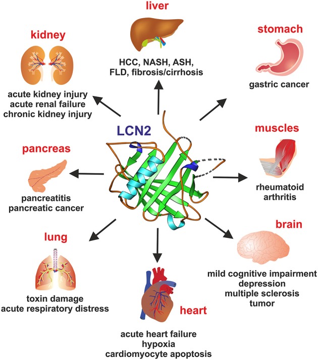
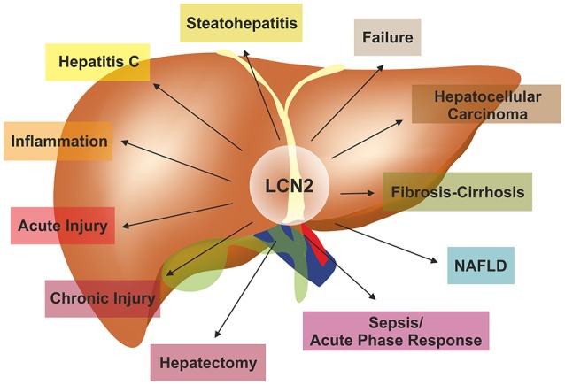
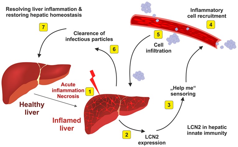
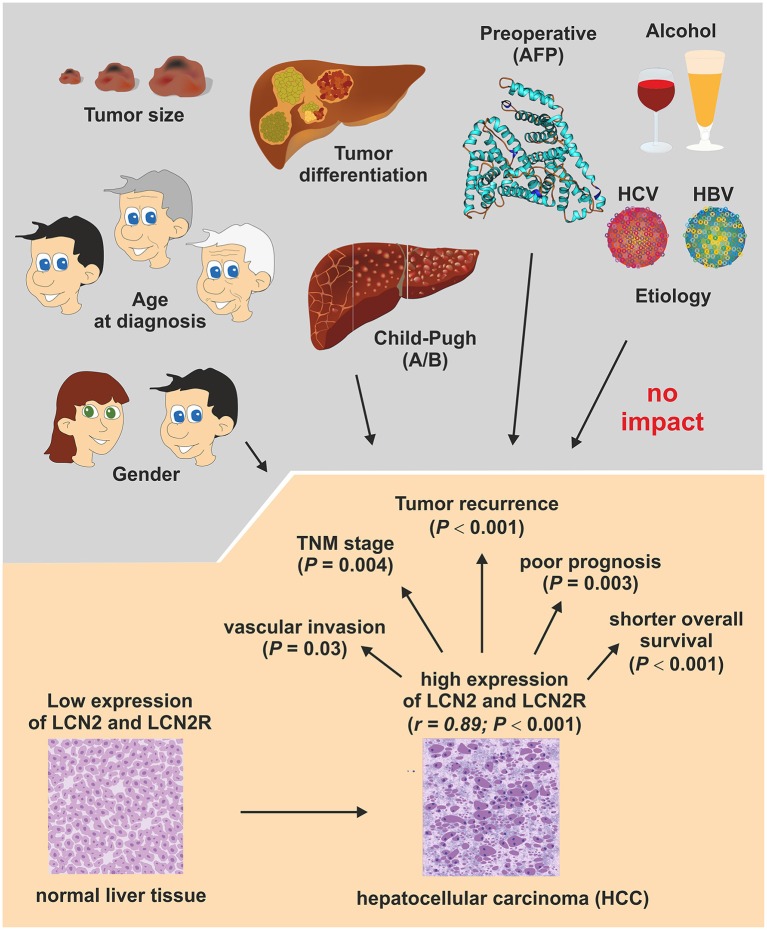
Lipocalin 2 (LCN2) is a secreted protein that belongs to the Lipocalins, a group of transporters of small lipophilic molecules such as steroids, lipopolysaccharides, iron, and fatty acids in circulation. Two decades after its discovery and after a high variety of published findings, LCN2's altered expression has been assigned to critical roles in several pathological organ conditions, including liver injury and steatosis, renal damage, brain injury, cardiomyopathies, muscle-skeletal disorders, lung infection, and cancer in several organs. The significance of this 25-kDa lipocalin molecule has been impressively increased during the last years. Data from several studies indicate the role of LCN2 in physiological conditions as well as in response to cellular stress and injury. LCN2 in the liver shows a protective role in acute and chronic injury models where its expression is highly elevated. Moreover, LCN2 expression is being considered as a potential strong biomarker for pathological conditions, including rheumatic diseases, cancer in human organs, hepatic steatosis, hepatic damage, and inflammation. In this review, we summarize experimental and clinical findings linking LCN2 to the pathogenesis of liver disease.

Keywords: alcoholic fatty liver disease; biomarker; hepatic disease; inflammation; liver; liver failure; matrix metalloproteinases; non-alcoholic steatohepatitis.


Figures

Figure 1
Figure 1
The lipocalin fold. Members of the lipocalin family have a typical eight-stranded, anti-parallel, symmetrical β-barrel fold structure. Depicted are human and mouse LCN2, human retniol-binding protein 4 (RBP4), and human liver fatty acid-binding protein (L-FABP). The depicted structures were generated using the Ribbons XP software (version 3.0) and coordinates 3BX8, 3S26, 3FMZ, and 2LKK deposited in the RCSB Protein Data Bank (http://www.rcsb.org). A size marker (10 Å) is given.
Figure 2
Figure 2
Lipocalin 2 (LCN2) in organ damage. Representative pathophysiological or malignancies conditions for which elevated LCN2 concentrations in representative organs are indicative are depicted.
Figure 3
Figure 3
LCN2 in the pathogenesis of liver diseases. LCN2 is a versatile adiponectin that influences all kinds of liver diseases. For more details on experimental and clinical findings associated with altered LCN2 expression refer to Table 3.
Figure 4
Figure 4
LCN2 and fat metabolism. A recent concept suggests that LCN2 is a key factor in controlling intracellular fat metabolisms in hepatocytes by regulating expression of the lipid droplet protein PLIN5/OXPAT. (A) The concept is majorly based on the finding that mice lacking LCN2 accumulate more lipids in the liver and show more hepatic damage and inflammation when fed a methionine-choline deficient diet (MCD) representing a nutritional model of NASH. (B) In the respective study, it was proposed that LCN2 imports lipids into hepatocytes either via specific receptors (e.g., LCN2R) or unknown endocytosis pathways. Within the cytoplasm, the LCN2/lipid complexes are first packed into endosomes whose slightly acidified microenvironment causes LCN2 to dissociate from the lipids. The lipids then move into the cytoplasm, where they are coated by PLIN5/OXPAT protecting them from intracellular degradation and oxidation. LCN2 may be recycled and secreted. PLIN5/OXPAT itself is up-regulated by PPAR-γ that is stimulated by the higher cytoplasmic fat content that is the consequence of facilitated lipid import by LCN2. Together, the suspected interaction of LCN2, PPAR-γ, and PLIN5/OXPAT presents a complex network that affects lipid metabolism, glucose homeostasis, and adipogenesis. In the presence of LCN2, intracellular concentrations of free reactive lipid species may be reduced and the overall inflammatory responses suppressed. Contrarily, in the absence of LCN2, the concentration of free fatty acids within the cytosol is increased predisposing for inflammation and steatosis.
Figure 5
Figure 5
Elevated hepatic LCN2 expression is a physiological indicator of hepatic inflammation. After acute inflammatory stimulus (1), the liver synthesizes and secretes large quantities of LCN2 (2). This physiological response indicates that the liver is inflamed and needs help (3). As a consequence of the emitted “help me” signal, inflammatory blood cells that are necessary to begin the repair are recruited (4) and infiltrate the liver (5). The infiltrating cell populations help to destroy and clear infectious particles (6) and are helpful in restoring tissue homeostasis (7). For more details about this hypothesis are given elsewhere (Asimakopoulou et al., 2016).
Figure 6
Figure 6
Association of LCN2 and LCN2R expression with conventional clinicopathological HCC parameters. An association study investigated the clinical significance of LCN2 and LCN2R in 138 patients who underwent curative resection of HCC. In conclusion, this study showed that the expression of both genes is correlated (r = 0.89; P < 0.001) and are up-regulated in HCC tissue and associated with vascular invasion status (P = 0.03) and TNM classification stage of malignant tumors (P = 0.004) that consider characteristics of the original primary tumor (T), the involved regional lymph nodes (N), and the occurrence of distant metastasis (M). In addition, the expression of both genes correlated well with tumor recurrence (P < 0.001), poor prognosis (P < 0.003), and overall survival rates (P < 0.001), suggesting that LCN2 and LCN2R expression are suitable prognostic factors and potential therapeutic targets in HCC. Details of this study are given elsewhere (Zhang et al., 2012).

References

    1. Aigner F., Maier H. T., Schwelberger H. G., Wallnöfer E. A., Amberger A., Obrist P., et al. . (2007). Lipocalin-2 regulates the inflammatory response during ischemia and reperfusion of the transplanted heart. Am. J. Transplant. 7, 779–788. 10.1111/j.1600-6143.2006.01723.x - DOI - PubMed
    1. Alwahsh S. M., Xu M., Seyhan H. A., Ahmad S., Mihm S., Ramadori G., et al. . (2014). Diet high in fructose leads to an overexpression of lipocalin-2 in rat fatty liver. World J. Gastroenterol. 20, 1807–1821. 10.3748/wjg.v20.i7.1807 - DOI - PMC - PubMed
    1. Ariza X., Graupera I., Coll M., Solà E., Barreto R., García E., et al. . (2016). Neutrophil gelatinase-associated lipocalin is a biomarker of acute-on-chronic liver failure and prognosis in cirrhosis. J. Hepatol. 65, 57–65. 10.1016/j.jhep.2016.03.002 - DOI - PubMed
    1. Ashraf M. I., Schwelberger H. G., Brendel K. A., Feurle J., Andrassy J., Kotsch K., et al. . (2016). Exogenous Lipocalin 2 ameliorates acute rejection in a mouse model of renal transplantation. Am. J. Transplant. 16, 808–820. 10.1111/ajt.13521 - DOI - PMC - PubMed
    1. Asimakopoulou A., Borkham-Kamphorst E., Henning M., Yagmur E., Gassler N., Liedtke C., et al. . (2014). Lipocalin-2 (LCN2) regulates PLIN5 expression and intracellular lipid droplet formation in the liver. Biochim. Biophys. Acta 1842, 1513–1524. 10.1016/j.bbalip.2014.07.017 - DOI - PubMed