. 2016 Sep 27:7:392.
doi: 10.3389/fimmu.2016.00392. eCollection 2016.

Utility of a Mouse Model of Osteoarthritis to Demonstrate Cartilage Protection by IFNγ-Primed Equine Mesenchymal Stem Cells

Affiliations

Utility of a Mouse Model of Osteoarthritis to Demonstrate Cartilage Protection by IFNγ-Primed Equine Mesenchymal Stem Cells

Marie Maumus et al. Front Immunol. .

Abstract

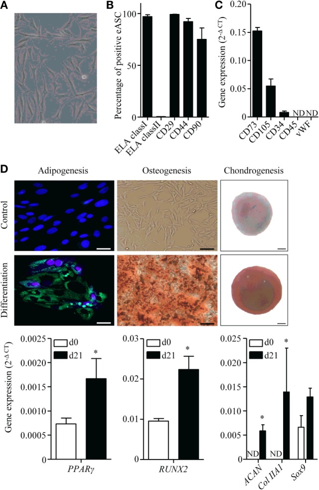
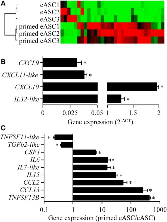
Objective: Mesenchymal stem cells isolated from adipose tissue (ASC) have been shown to influence the course of osteoarthritis (OA) in different animal models and are promising in veterinary medicine for horses involved in competitive sport. The aim of this study was to characterize equine ASCs (eASCs) and investigate the role of interferon-gamma (IFNγ)-priming on their therapeutic effect in a murine model of OA, which could be relevant to equine OA.

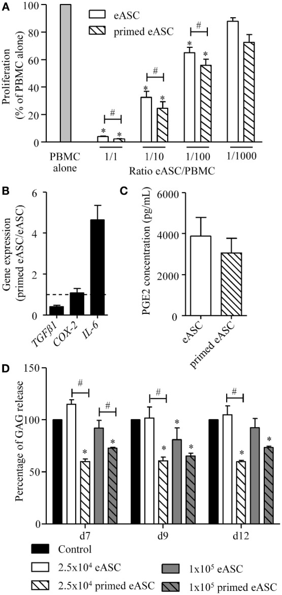
Methods: ASC were isolated from subcutaneous fat. Expression of specific markers was tested by cytometry and RT-qPCR. Differentiation potential was evaluated by histology and RT-qPCR. For functional assays, naïve or IFNγ-primed eASCs were cocultured with peripheral blood mononuclear cells or articular cartilage explants. Finally, the therapeutic effect of eASCs was tested in the model of collagenase-induced OA (CIOA) in mice.

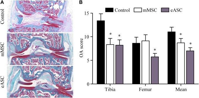
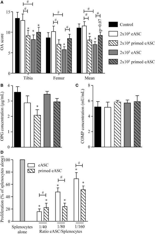
Results: The immunosuppressive function of eASCs on equine T cell proliferation and their chondroprotective effect on equine cartilage explants were demonstrated in vitro. Both cartilage degradation and T cell activation were reduced by naïve and IFNγ-primed eASCs, but IFNγ-priming enhanced these functions. In CIOA, intra-articular injection of eASCs prevented articular cartilage from degradation and IFNγ-primed eASCs were more potent than naïve cells. This effect was related to the modulation of eASC secretome by IFNγ-priming.

Conclusion: IFNγ-priming of eASCs potentiated their antiproliferative and chondroprotective functions. We demonstrated that the immunocompetent mouse model of CIOA was relevant to test the therapeutic efficacy of xenogeneic eASCs for OA and confirmed that IFNγ-primed eASCs may have a therapeutic value for musculoskeletal diseases in veterinary medicine.

Keywords: cartilage; cell therapy; horse; mesenchymal stem cells; osteoarthritis; secretome.


Figures

Figure 1
Figure 1
Characterization of equine ASCs. (A) Representative photomicrograph of eASCs. (B) Percentage of eASCs positive for the indicated membrane markers. Results are percentage of positive cells and are expressed as mean ± SEM for three separate experiments. (C) Gene expression level of indicated markers in eASCs. Results are relative expression (2−ΔCT) and are expressed as mean ± SEM for three separate experiments. (D) Differentiation of eASCs: adipogenesis is characterized by the expression of peroxysome proliferator-activated receptor (PPAR)-γ at day 21 (d21) versus d0 and by the visualization of lipid droplets stained with HCS LipidTOX™ Green neutral lipid stain and counterstained with DAPI (lower panel) versus control proliferative medium (upper panel) at d21 (scale bar is 25 μm). Osteogenesis is characterized by the expression of Runx2 at d21 versus d0 and by Alizarin Red S positive staining in differentiation versus proliferative medium at d21 (scale bar is 100 μm). Chondrogenesis is characterized by the expression of Sox9, collagen IIA1 (col IIA1), and aggrecan (Agg) at d21 versus d0 and by Safranin O staining (lower panel) versus undifferentiated control (upper panel) at d21 (scale bar is 100 μm). Results are expressed as relative expression (2−ΔCT) and represented as mean ± SEM for three to six independent biological replicates. Data were analyzed using the Mann–Whitney test. *p < 0.05. ND: not detected.
Figure 2
Figure 2
IFNγ-priming improves the immunosuppressive and chondroprotective properties of eASCs in vitro. (A) Proliferation of equine PBMCs in presence of naïve eASCs or IFNγ-primed eASCs at different ratios. Results are expressed as the percentage of PHA-induced proliferation of equine PBMCs, which was assigned the value of 100% and represented as mean ± SEM for three independent biological replicates. (B) Gene expression level of TGFβ1, COX-2, and IL-6 in eASCs. Gene expression in IFNγ-primed eASCs was normalized to that obtained in naïve eASCs. (C) PGE2 concentration in supernatants of naïve and IFNγ-primed eASCs. (D) GAG release quantification from cartilage explants incubated with IL1β (1 ng/mL) and Oncostatin M (10 ng/mL) during 7, 9, or 12 days and cocultured with or without 25,000 or 100,000 naïve and IFNγ-primed eASCs (100 ng/mL, 24 h before coculture). Results are expressed as the percentage of GAG release normalized to control condition (explant alone) at each time points and represented as mean ± SEM for 10–14 independent biological replicates. Data were analyzed using the Kruskal–Wallis test followed by Dunn’s test for multiple comparisons for (A, D) and using the Mann–Whitney test for (B,C) *p < 0.05 in samples versus PBMC alone (A) or control (D); #p < 0.05 between primed and unprimed samples.
Figure 3
Figure 3
Xenogeneic eASCs improve OA scores in the collagenase-induced OA mouse model. OA was induced by intra-articular injection of collagenase in mouse knee joints and the severity of OA was evaluated at day 42. (A) Representative photographs of knee joints from control (collagenase alone) (upper), mMSCs-treated (middle), and eASCs-treated (lower) mice. (B) Histological OA score of tibia plateaus and femur condyles in the mouse knee joints and mean at euthanasia. Results are expressed as the mean ± SEM, n = 10. Data were analyzed using the Kruskal–Wallis test followed by Dunn’s test for multiple comparisons. *p < 0.05.
Figure 4
Figure 4
IFNγ-primed eASCs improve OA score. (A) OA score for tibia plateaus and femur condyles in the knee joint and mean at euthanasia. Results are expressed as the mean ± SEM, n = 10. (B) Osteoprotegerin (OPG) and (C) cartilage oligomeric matrix protein (COMP) concentrations were determined in sera of mice at euthanasia by specific sandwich enzyme-linked immunosorbent assay (ELISA). Results are expressed as the mean ± SEM, n = 10. (D) Proliferation of murine splenocytes in presence of different ratios of naïve or IFNγ-primed eASCs. Results are expressed as the percentage of ConA-induced proliferation of splenocytes which was assigned the value of 100% and represented as mean ± SEM for three independent biological replicates. Data were analyzed using the Kruskal–Wallis test followed by Dunn’s test for multiple comparisons in A, B, and C and using ANOVA followed by a Dunnett’s test for multiple comparisons in D. *p < 0.05 or p = 0.07 compared samples with the control; #p < 0.05 compared samples with each other.
Figure 5
Figure 5
Effect of IFNγ-priming on the inflammatory gene profile of eASCs. Gene array analysis of inflammatory cytokines and chemokines mRNA compared naïve and IFNγ-primed eASCs. (A) Hierarchical clustering comparing naïve or IFNγ-primed eASCs, (B) induced gene expression levels in IFNγ-primed eASCs expressed as relative expression (2−ΔCT), (C) significantly modulated gene expression levels in IFNγ-primed eASCs. Results are represented as mean ± SEM for three independent biological replicates. Data were analyzed using the Mann–Whitney test. *p < 0.05.

References

    1. Goodrich LR, Nixon AJ. Medical treatment of osteoarthritis in the horse – a review. Vet J (2006) 171(1):51–69.10.1016/j.tvjl.2004.07.008 - DOI - PubMed
    1. Maninchedda U, Lepage OM, Gangl M, Hilairet S, Remandet B, Meot F, et al. Development of an equine groove model to induce metacarpophalangeal osteoarthritis: a pilot study on 6 horses. PLoS One (2015) 10(2):e0115089.10.1371/journal.pone.0115089 - DOI - PMC - PubMed
    1. Whitworth DJ, Banks TA. Stem cell therapies for treating osteoarthritis: prescient or premature? Vet J (2014) 202(3):416–24.10.1016/j.tvjl.2014.09.024 - DOI - PubMed
    1. Schnabel LV, Fortier LA, McIlwraith CW, Nobert KM. Therapeutic use of stem cells in horses: which type, how, and when? Vet J (2013) 197(3):570–7.10.1016/j.tvjl.2013.04.018 - DOI - PubMed
    1. Vidal MA, Kilroy GE, Lopez MJ, Johnson JR, Moore RM, Gimble JM. Characterization of equine adipose tissue-derived stromal cells: adipogenic and osteogenic capacity and comparison with bone marrow-derived mesenchymal stromal cells. Vet Surg (2007) 36(7):613–22.10.1111/j.1532-950X.2007.00313.x - DOI - PubMed