. 2016 Oct 12:6:35103.
doi: 10.1038/srep35103.

Riboflavin photoactivation by upconversion nanoparticles for cancer treatment

Affiliations

Riboflavin photoactivation by upconversion nanoparticles for cancer treatment

E V Khaydukov et al. Sci Rep. .

Abstract

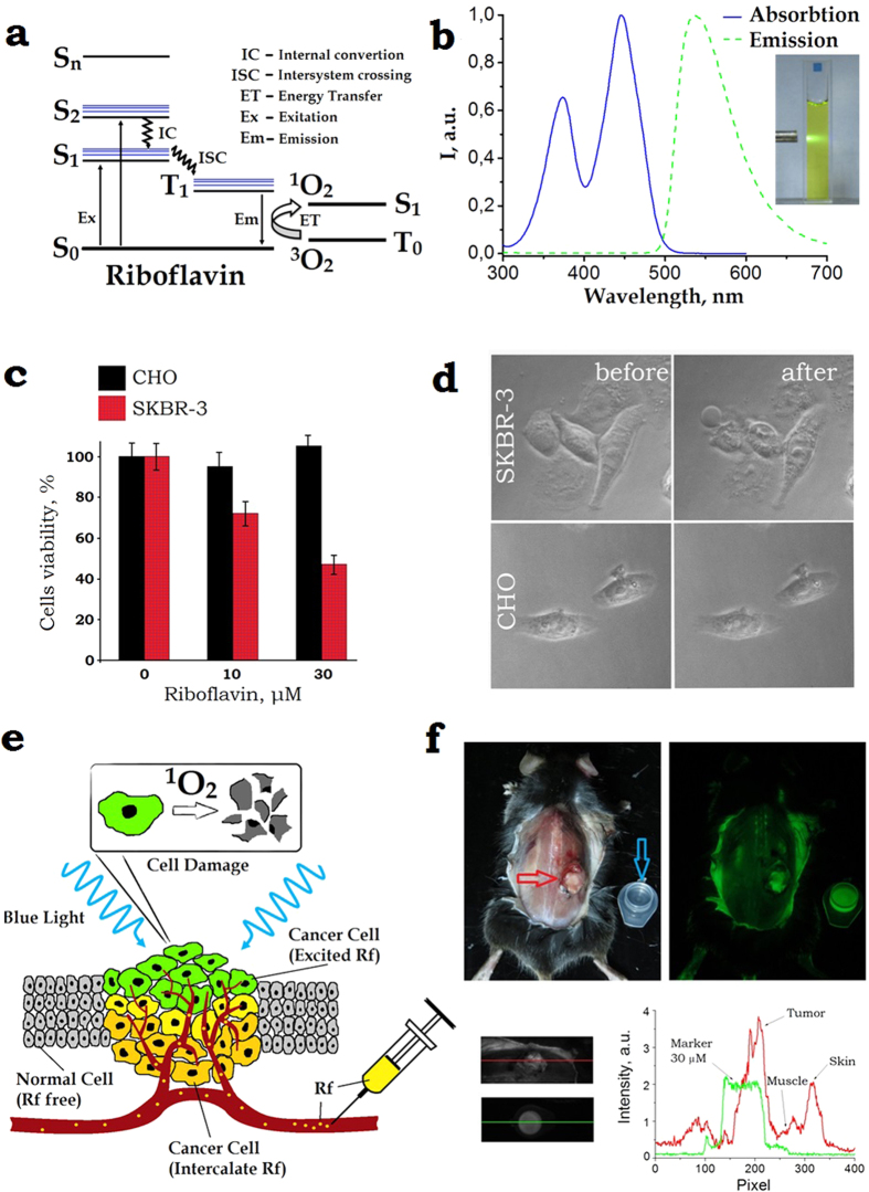
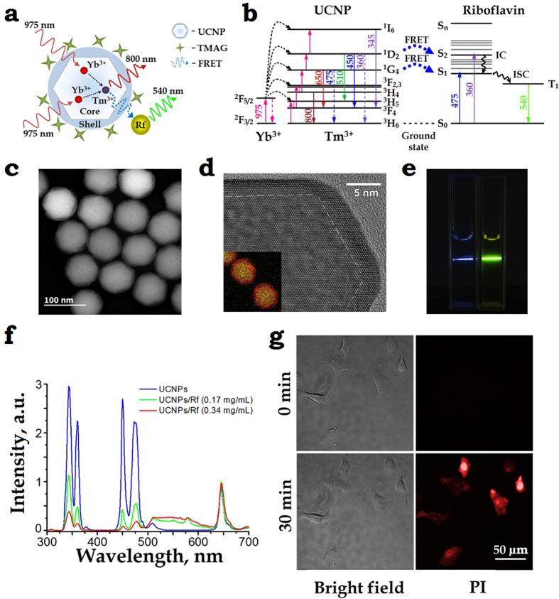
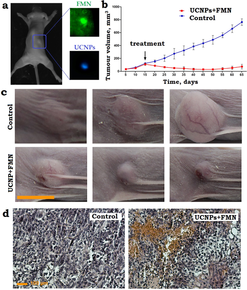
Riboflavin (Rf) is a vitamin and endogenous photosensitizer capable to generate reactive oxygen species (ROS) under UV-blue irradiation and kill cancer cells, which are characterized by the enhanced uptake of Rf. We confirmed its phototoxicity on human breast adenocarcinoma cells SK-BR-3 preincubated with 30-μM Rf and irradiated with ultraviolet light, and proved that such Rf concentrations (60 μM) are attainable in vivo in tumour site by systemic intravascular injection. In order to extend the Rf photosensitization depth in cancer tissue to 6 mm in depth, we purpose-designed core/shell upconversion nanoparticles (UCNPs, NaYF4:Yb3+:Tm3+/NaYF4) capable to convert 2% of the deeply-penetrating excitation at 975 nm to ultraviolet-blue power. This power was expended to photosensitise Rf and kill SK-BR-3 cells preincubated with UCNPs and Rf, where the UCNP-Rf energy transfer was photon-mediated with ~14% Förster process contribution. SK-BR-3 xenograft regression in mice was observed for 50 days, following the Rf-UCNPs peritumoural injection and near-infrared light photodynamic treatment of the lesions.


Figures

Figure 1
Figure 1
(a) Schematic diagram of the energy levels of riboflavin. A photon excites Rf from the singlet ground state (S0) to the first excited singlet state (S1) from which it can either decay to the S0 or undergo intersystem crossing to an excited triplet state (T1). Rf in the long-lived T1 state is capable to non-radiatively (collisionally) drive environmental oxygen from its triplet ground state 3O2 to the chemically reactive singlet excited state 1O2. The excitation and emission spectra of Rf are shown in Panel (b). (c) The MTT assay of the cells after incubation with Rf for 90 min followed by 365-nm light exposure at the dose 4.2 J/cm2. SK-BR-3 and CHO cells are marked red and black, respectively. The riboflavin CRf ≈ 30 μM reduced the SKBR-3 cells viability to 47 ± 7%. Error bars represent standard deviation (SD) for three independent experiments. (d) Phase-contrast images of SK-BR-3 and CHO cells treated with 30-μM riboflavin, before (left) and 2-h after (right) 10-min 365-nm light irradiation. Rapture of SK-BR-3 cells membrane was evident (bubbles), whereas the control CHO cells showed no morphological changes. (e) Schematic representation of the Rf-aided photodynamic treatment of superficial layers of targeted cancer cells shown as polymorphic yellow/green-coloured cells embedded into a layer of monomorphic grey-coloured normal cells. Buffer solution of Rf is injected into the tumour interstitium. 1O2 generated in the superficial cancer cell layer (green-coloured) exposed to UV-blue light induce phototoxicity. (f) Top row: left and right panels, post mortem bright-field and epi-luminescent images of BDF1 mice with a Lewis lung carcinoma tumour grafted on the dorsal side (marked by a red arrow), respectively. A plastic cuvette filled with CRf ≈ 30 μM is shown besides the animal (marked by a blue arrow). The epi-luminescent image was acquired under 450-nm excitation; fluorescence detection spectral band was 500–570 nm. Bottom row: bright-field zoomed images of the grafted tumour and cuvette and its fluorescence signal intensity profiles.
Figure 2
Figure 2
(a) A schematic diagram of the core/shell UCNP, explaining the 975-nm excitation (solid red wavy lines) of Yb3+ ions in the core, which then non-radiatively transfers (dashed arrows) the energy to an Tm3+ ion that passes the energy (dashed blue wavy line) to an Rf molecule via a resonant energy transfer (RET) process. The UCNP surface is coordinated by tetramethylammonium hydroxide (TMAH). (b) Energy level diagram of a UCNP – Rf pair. Excitation at 975 nm drives Yb3+ to the excited state 2F5/2 from which it can non-radiatively transfer the energy to Tm3+ (via 3H6 → 3H5 transition, followed by the relaxation to the metastable level 3F4). Two metastable excited state 2F5/2 Yb3+ and 3F4 Tm3+ ions coalesce to drive Tm3+ to the 3F2,3 state, while an Yb3+ decays to the ground state 2F3/2. Likewise, collective energy process of 2F5/2 Yb3+ and 3F4. 3H4. 1G4 … Tm3+ drives Tm3+ to the 3H4.1G4. 1D2 …, respectively (magenta arrows). There is a probability to populate S1, S2 levels of Rf via RET or Föster RET processes from the 1G4, 1D2 levels of Tm3+. (c) High-angle annular dark-field scanning and (d) high-resolution TEM images of as-synthesised core/shell NaYF4:Yb:Tm/NaYF4 UCNPs mean-sized 75 ± 5 nm, featuring the β-crystal phase. Overlay Y (green) and Yb (red) elemental EDX mapping of UCNPs is given as a bottom left insert in (d). (e) In vitro demonstration of RET of a UCNP–Rf donor-acceptor pair. Two cuvettes filled with plain UCNPs and UCNP-FMN 0.34 mg/mL aqueous colloids illuminated with a 975-nm laser beam. The respective blue and yellow traces of photoluminescence illustrate a strong RET effect. (f) Spectra of UCNP- FMN in water under 975-nm excitation acquired at 0, 0.17 mg/mL and 0.34 mg/mL concentrations of Rf (blue, green, red curves), respectively, with the concentration of UCNPs 0.5 mg/mL. A broadband fluorescence signal from 500 nm to 620 nm ascribed to the FMN emission was a strong manifestation of the RET between UCNP and FMN. (g) Demonstration of the phototoxicity effect of UCNP-Rf pair on SK-BR-3 cells irradiated with a 975-nm laser. Phase contrast (left) and propidium iodide (PI) fluorescence (right) images of the cells before (top images) as compared to the cells after (bottom image) irradiation highlights disintegration of the cells membranes.
Figure 3
Figure 3
(a) A photograph of the dorsal side of the immunodeficient mouse, bearing a grafted subcutaneously SK-BR-3 tumour 15 days post-implantation. A PBS solution of FMN and UCNPs was injected peritumourally. The tumour area is marked by a (blue) rectangle, its zoomed-in images spectrally filtered to emphasise FMN and UCNPs emissions are shown in insets labelled FMN and UCNPs, respectively. In FMN and UCNP images, the tumour exhibited contrast of 2 and 30, respectively, demonstrating the superiority of UCNP-assisted imaging. (b) A plot of the SK-BR-3 tumour evolution, showing progressive stable growth of the control tumours (non-irradiated) and tumour regression post-PDT treatment (black arrow, day 15) using FMN + UCNPs. (c) A time-lapse series of the bright-field photographs of the SK-BR-3 tumour area taken prior to the 975-nm laser treatment “15”, 25 and 50 days after treatment and the photographs of appropriate controls. Scale bar, 10 mm. (d) Histological images of the tumour tissue sections stained with hematoxylin and eosin, excised 1 day after the 975-nm PDT treatment. FMN + UCNPs in PBS solution were injected peritumourally in the control and PDT treated tumours. FMN + UCNPs after irradiation display profound hemorrhages, respectively, whereas control shows no abnormalities. Scale bar, 100 μm.

References

    1. Morris H. P. Effects on the genesis and growth of tumors associated with vitamin intake. Annals of the New York Academy of Sciences 49, 119–140, doi: 10.1111/j.1749-6632.1947.tb30939.x (1947). - DOI
    1. Rao P. N. et al.. Elevation of serum riboflavin carrier protein in breast cancer. Cancer Epidemiology Biomarkers & Prevention 8, 985–990 (1999). - PubMed
    1. Bareford L. M., Phelps M. A., Foraker A. B. & Swaan P. W. Intracellular processing of riboflavin in human breast cancer cells. Molecular Pharmaceutics 5, 839–848, doi: 10.1021/mp800046m (2008). - DOI - PubMed
    1. Bareford L. M., Avaritt B. R., Ghandehari H., Nan A. & Swaan P. W. Riboflavin-Targeted Polymer Conjugates for Breast Tumor Delivery. Pharmaceutical Research 30, 1799–1812, doi: 10.1007/s11095-013-1024-5 (2013). - DOI - PubMed
    1. Conrad K. S., Bilwes A. M. & Crane B. R. Light-Induced Subunit Dissociation by a Light-Oxygen-Voltage Domain Photoreceptor from Rhodobacter sphaeroides. Biochemistry 52, 378–391, doi: 10.1021/bi3015373 (2013). - DOI - PMC - PubMed

Publication types

MeSH terms

LinkOut - more resources