. 2016 Oct 12:6:35263.
doi: 10.1038/srep35263.

New potential beneficial effects of actein, a triterpene glycoside isolated from Cimicifuga species, in breast cancer treatment

Affiliations

New potential beneficial effects of actein, a triterpene glycoside isolated from Cimicifuga species, in breast cancer treatment

Grace Gar-Lee Yue et al. Sci Rep. .

Abstract

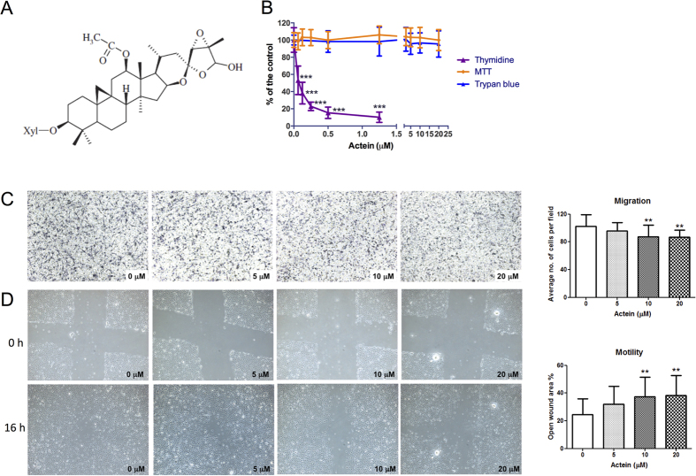
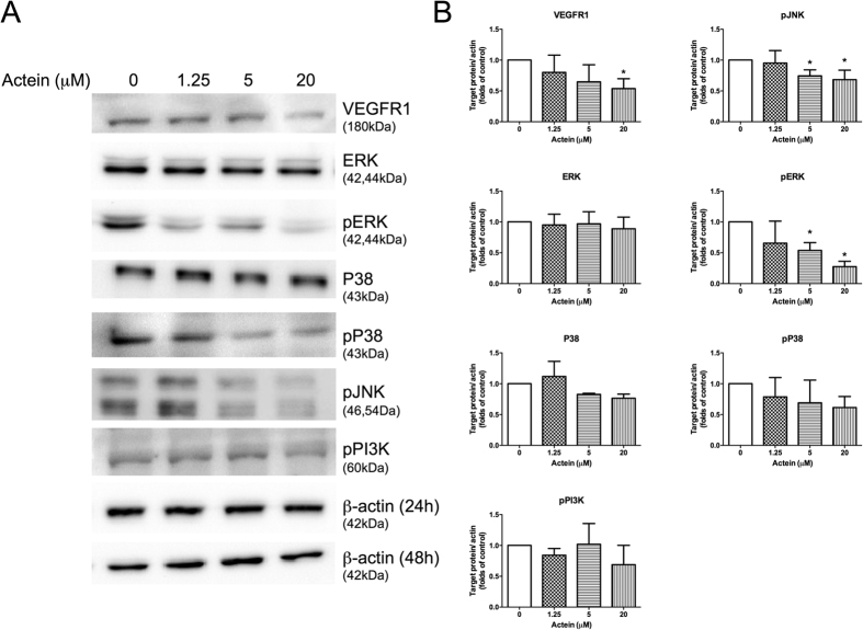
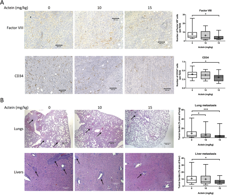
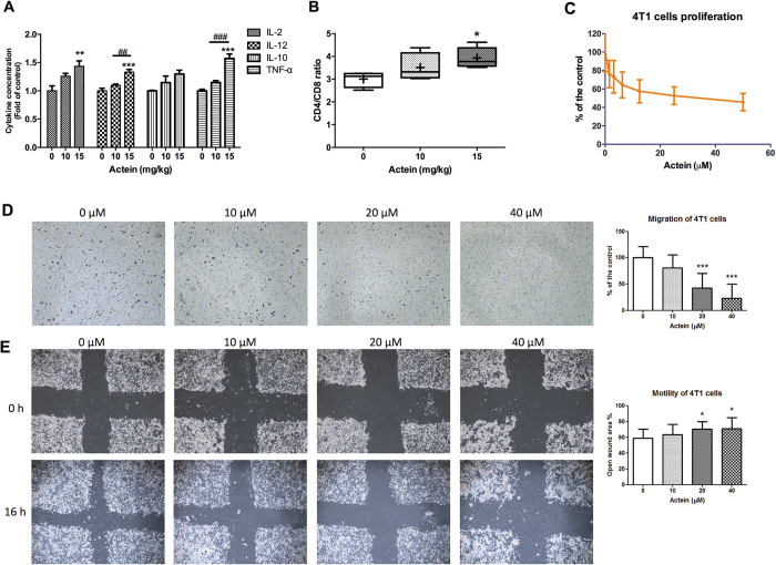
Actein is a triterpene glycoside isolated from the rhizomes of Cimicifuga foetida (Chinese herb "shengma") which could inhibit the growth of breast cancer cells. Nevertheless, the effect of actein on angiogenesis, which is an essential step for tumor growth and metastasis, has never been reported. Hence, this study aimed to investigate the in vitro and in vivo effects of actein on angiogenesis using human microvascular endothelial cells (HMEC-1), matrigel plug and tumor-bearing mouse models. Our results showed that actein significantly inhibited the proliferation, reduced the migration and motility of endothelial cells, and it could suppress the protein expressions of VEGFR1, pJNK and pERK, suggesting that JNK/ERK pathways were involved. In vivo results showed that oral administration of actein at 10 mg/kg for 7 days inhibited blood vessel formation in the growth factor-containing matrigel plugs. Oral actein treatments (10-15 mg/kg) for 28 days resulted in decreasing mouse 4T1 breast tumor sizes and metastasis to lungs and livers. The apparent reduced angiogenic proteins (CD34 and Factor VIII) expressions and down-regulated metastasis-related VEGFR1 and CXCR4 gene expressions were observed in breast tumors. Our novel findings provide insights into the use of actein for development of anti-angiogenic agents for breast cancer.


Figures

Figure 1
Figure 1. Effects of actein on cell proliferation, migration and motility of HMEC-1 cells.
(A) Chemical structure of actein. (B) Cells were treated with different concentrations of actein for 48 hours, then cytotoxicity and cell proliferation was determined by MTT, trypan blue and [methyl-3H] thymidine incorporation assays, respectively. Results are expressed as the mean % ratio of optical density or count per minute in treated and vehicle-treated control wells (mean ± SD of 3 independent experiments with 5 wells each). (C) Representative photomicrographs showing the migrated and stained HMEC-1 cells on the lower side of membranes in the presence or absence of actein. Quantification of cell migration in modified Boyden chambers was shown. Results are expressed in number of cells per field (mean + SD of 4 independent experiments). (D) Representative photomicrographs showing the cells migrated across the scratch wound in the presence or absence of actein after 16 hours of incubation. Quantification of wound-induced cell motility in HMEC-1 was shown. Results are expressed as the mean open wound area (mean + SD of 3 independent experiments). Differences among the treated and vehicle-treated control groups were determined by one-way ANOVA with Tukey’s post-hoc test. **p < 0.01, ***p < 0.001 as compared to control group.
Figure 2
Figure 2. Effects of actein on expressions of VEGFR1 and signaling kinases in HMEC-1 cells.
Western blot analyses on P38, pP38, ERK, pERK, pJNK, pPI3K expressions in actein-treated HMEC-1 cells for 24 hours (or 48 hours for VEGFR1) were performed. (A) Immunoblotting was performed 3 times using independently prepared cell lysates and the representative blots were shown. (B) The histograms showed the quantified results of protein levels, which were adjusted with corresponding β-actin protein levels and expressed as fold of control (mean fold of control + SD from 3 independent experiments). Differences among the treated and vehicle-treated control groups were determined by one-way ANOVA with Tukey’s post-hoc test. *p < 0.05 as compared to control group.
Figure 3
Figure 3. Effects of actein on angiogenesis and tumor growth in mouse models.
(A) Upper: Hemoglobin content of matrigel plugs from different treatment groups (n = 9-10). Lower: The photos of matrigel plugs excised from mice of different treatment groups 7 days after inoculation. (B–E) 4T1 tumor-bearing mice were treated with actein (10 or 15 mg/kg) or vehicle for 4 weeks. The volumes of tumors (B) and body weights (C) were recorded and expressed as mean ± SEM (n = 12). (D) The final tumor weights after actein or vehicle treatments. (E) Quantitative RT-PCR analyses of VEGFRs, Ang1, Ang2, Tie1, CXCR4, AKT mRNA expressions in tumors excised from different treatment groups. Tumor tissues were collected for RNA extraction. Data were normalized to corresponding human beta-2 microglobulin (B2M) expressions in tumors of vehicle-treated group. mRNA expressions results are expressed as fold of control (mean fold of control + SEM from 8 mice each group). Differences among the treated and vehicle-treated control groups were determined by one-way ANOVA with Tukey’s post-hoc test. *p < 0.05, **p < 0.01, ***p < 0.001 as compared to control group.
Figure 4
Figure 4. Histopathology of tumors, lungs and livers of tumor-bearing mice after actein treatments.
(A) Endothelial cells in the tumor sections were assessed using Factor VIII and CD34 immunohistochemical analysis. Representative photomicrographs of magnification 40X showing the endothelial cells stained with anti-Factor VIII or anti-CD34 antibodies in brown. Quantification of Factor-VIII or CD34 positive-stained cells in tumor sections was conducted in a blinded manner. Results were expressed in box and whisker plots of 12 mice each group. (B) The paraffin-embedded sections of the lungs and livers were photographed and used to measure metastatic loci area and total lung or liver area. The histograms showed the tumor burden in lungs and livers according to the tumor area as a percentage of whole lung or liver area per group. Representative H&E-stained sections of lungs and livers from different groups with arrows showing the metastatic loci. Results were expressed in box and whisker plots of 12 mice each group. Differences among the treated and vehicle-treated control groups were determined by one-way ANOVA with Tukey’s post-hoc test. *p < 0.05, **p < 0.01, ***p < 0.001 as compared to control group.
Figure 5
Figure 5. Effects of actein on cytokine profiles and T-lymphocyte subset distribution of tumor-bearing mice and on 4T1 cell proliferation and migration.
Spleens of tumor-bearing mice were excised for lymphocyte isolation as described in methodology section. Lymphocytes were incubated for 24 hours and the culture supernatants were collected. Cytokines concentrations of (A) IL-2, IL-10, IL-12, TNF-α were specifically determined by ELISA. Results were expressed as fold of control (mean fold of control + SEM from 6 mice each group). Another portion of spleen lymphocytes were with T-lymphocyte subset antibody cocktail and the number of stained cells for CD3, CD4 and CD8 were quantified by fluorescence activated cell sorting and were depicted here as the ratio of CD4+ and CD8+ cells per 20,000 viable cells. (B) Results were expressed in box and whisker plots of 6 mice each group. (C) 4T1 cells were treated with different concentrations of actein for 48 hours, then cell proliferation was determined by [methyl-3H] thymidine incorporation assays. Results are expressed as the mean % count per minute in treated and vehicle-treated control wells (mean ± SD of 3 independent experiments with 5 wells each). (D) Representative photomicrographs showing the migrated and stained 4T1 cells on the lower side of membranes in the presence or absence of actein. Quantification of cell migration in modified Boyden chambers was shown. Results are expressed in number of cells per field (mean + SD of 4 independent experiments). (E) Representative photomicrographs showing the cells migrated across the scratch wound in the presence or absence of actein after 16 hours of incubation. Quantification of wound-induced cell motility in 4T1 was shown. Results are expressed as the mean open wound area (mean + SD of 3 independent experiments). Differences among the treated and vehicle-treated control groups were determined by one-way ANOVA with Tukey’s post-hoc test. **p < 0.01, ***p < 0.001 as compared to control group. *p < 0.05, **p < 0.01, ***p < 0.001 as compared to control group. ##p < 0.01, ###p < 0.001 as compared among groups.

References

    1. McKenna D. J., Jones K., Humphrey S. & Hughes K. Black cohosh: efficacy, safety, and use in clinical and preclinical applications. Altern. Ther. Health Med. 7, 93–100 (2001). - PubMed
    1. Li J. X. & Yu Z. Y. Cimicifugae rhizoma: from origins, bioactive constituents to clinical outcomes. Curr. Med. Chem. 13, 2927–2951 (2006). - PubMed
    1. Sakurai N. et al. Cancer preventive agents. Part 1: chemopreventive potential of cimigenol, cimigenol-3,15-dione, and related compounds. Bioorg. Med. Chem. 13, 1403–1408 (2005). - PubMed
    1. Commission C. P. Chinese Pharmacopoeia. 73–74 (China Medical Science and Technology Press, 2015).
    1. Einbond L. S. Black cohosh: chemopreventive and anticancer potential. In: Complementary and alternative therapies and the aging population: an evidence-based approach. (ed. Watson R. R. ) 193–227 (Elsevier Academic Press, 2009).

Publication types