Review
. 2016 Dec;142(12):1275-1314.
doi: 10.1037/bul0000073. Epub 2016 Oct 10.

Dispositional negativity: An integrative psychological and neurobiological perspective

Affiliations
Review

Dispositional negativity: An integrative psychological and neurobiological perspective

Alexander J Shackman et al. Psychol Bull. 2016 Dec.

Abstract

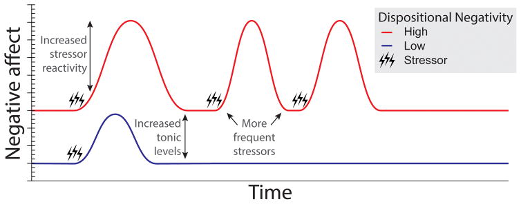
Dispositional negativity-the propensity to experience and express more frequent, intense, or enduring negative affect-is a fundamental dimension of childhood temperament and adult personality. Elevated levels of dispositional negativity can have profound consequences for health, wealth, and happiness, drawing the attention of clinicians, researchers, and policymakers. Here, we highlight recent advances in our understanding of the psychological and neurobiological processes linking stable individual differences in dispositional negativity to momentary emotional states. Self-report data suggest that 3 key pathways-increased stressor reactivity, tonic increases in negative affect, and increased stressor exposure-explain most of the heightened negative affect that characterizes individuals with a more negative disposition. Of these 3 pathways, tonically elevated, indiscriminate negative affect appears to be most central to daily life and most relevant to the development of psychopathology. New behavioral and biological data provide insights into the neural systems underlying these 3 pathways and motivate the hypothesis that seemingly "tonic" increases in negative affect may actually reflect increased reactivity to stressors that are remote, uncertain, or diffuse. Research focused on humans, monkeys, and rodents suggests that this indiscriminate negative affect reflects trait-like variation in the activity and connectivity of several key brain regions, including the central extended amygdala and parts of the prefrontal cortex. Collectively, these observations provide an integrative psychobiological framework for understanding the dynamic cascade of processes that bind emotional traits to emotional states and, ultimately, to emotional disorders and other kinds of adverse outcomes. (PsycINFO Database Record


Conflict of interest statement

Authors declare no conflicts of interest.

Figures

Figure 1
Figure 1. Pathways linking dispositional negativity (trait) to increased momentary negative affect (state)
Questionnaire and behavioral data suggest that three key pathways—increased stressor reactivity, increased tonic levels of negative affect, and more frequent stressors—explain most of the heightened negative affect characteristic of individuals with a negative disposition. Lines depict hypothesized fluctuations in momentary negative affect in individuals with high (red) and low (blue) levels of dispositional negativity, respectively. Acute stressors (e.g., daily hassles, social conflict, and negative life events) are indicated by black lightning bolts.
Figure 2
Figure 2. Different kinds of threat
A. Rodents (Green). In rats and mice, the open field test and the elevated plus maze (EPM) are commonly used to assess emotional responses to diffuse threat. In the open field, rodents are placed into a relatively large, brightly lit, and unfamiliar context. In the elevated plus maze, rodents are placed in a maze with two open arms and two arms enclosed by walls. Freezing and avoidance of the center of the open field or the open arms of the maze provide behavioral ‘read-outs’ of negative affect. Figure adapted with permission from (Tovote et al., 2015). B. Nonhuman primates (Orange). In monkeys, the Human ‘Intruder’ Paradigm (HIP) can be used to quantify naturalistic defensive behaviors, neuroendocrine activity, and brain metabolic activity associated with exposure to a range of threats. In the ‘Alone’ (ALN) condition, the monkey is simply placed in the testing cage. This novel, diffusely threatening context elicits low levels of freezing and cortisol and a moderate frequency of alarm and separation calls. In the ‘No Eye Contact’ (NEC) condition (A, left), the intruder presents his or her profile while avoiding making eye contact. This elicits passive defenses, including freezing and vocal reductions, similar to procedures used for quantifying behavioral inhibition in children. Panel adapted with permission from (A. S. Fox & Kalin, 2014). C. Humans (Purple). In humans, a wide variety of paradigms have been used to probe responses to uncertain, diffuse, or remote threat. Often, these involve the unpredictable presentation of electric shocks or, as shown in the accompanying figure, aversive images. For example, Somerville and colleagues assessed neural activity associated with the temporally certain or uncertain presentation of aversive images. Figure adapted with permission from (Somerville et al., 2013). D. Comparison of paradigms (Scatter plot). Threats differ along several key dimensions, including certainty (x-axis), physical or temporal imminence, diffuseness (y-axis; specific cues vs. real or virtual contexts), and duration (dot size). Here we present some common paradigms used in rodents (green), nonhuman primates (organge), and humans (purple). Studies were chosen for illustrative purposes. We did not attempt a comprehensive review of the literature and, of necessity, the locations of particular paradigms along the two dimensions of the scatter plot are approximate and somewhat arbitrary. Interestingly, many paradigms confound multiple dimensions (e.g., if vs. when threat will occur) and, because of temporal constraints imposed by conventional fMRI techniques, human imaging studies have focused on the relatively brief (<2 min) anticipation of uncertain threat. Studies [duration in sec.]—Rodents: Alone in brightly lit cage [900] (D. L. Walker & Davis, 1997); CS+, 100% probability [30] (Duvarci et al., 2009); CS−, 0% probability [30] (Duvarci et al., 2009); Elevated plus maze (EPM) [900] (Tye et al., 2011); Open field [1080] (Tye et al., 2011); Temporally unpredictable cued shock [mean=162] (Miles, Davis, & Walker, 2011); Unpredictable shock context [1200] (Luyten et al., 2012). Monkeys: Alone (ALN) [1800] (A. S. Fox et al., 2008); CS+, 100% probability [4] (Winslow, Noble, & Davis, 2007); CS−, 0% probability [10] (Kalin, Shelton, Davidson, & Lynn, 1996); No eye contact (NEC) [1800] (A. S. Fox et al., 2008). Humans: Alone in dark room [120] (Grillon, Pellowski, Merikangas, & Davis, 1997); Anticipating image with unpredictable valence [5] (Grupe et al., 2013); CS+, 100% probability [8] (Gazendam et al., 2013); CS+, 50% probability [3] (Buchel, Morris, Dolan, & Friston, 1998); CS−, 0% shock probability (conditioned safety cue) [8] (Gazendam et al., 2013); Tarantula (video clip of approach) [4] (Mobbs et al., 2010); Temporally unpredictable aversive image [115] (Somerville et al., 2013); Temporally unpredictable cued shock [mean=140] (Moberg & Curtin, 2009); Trier Social Stress Test (TSST) [780] (http://topics.sciencedirect.com/topics/page/Trier_social_stress_test); Virtual reality context paired with unpredictable shock [40] (Alvarez, Chen, Bodurka, Kaplan, & Grillon, 2011).
Figure 3
Figure 3. Central extended amygdala circuitry
Simplified schematic of key inputs and outputs to the central extended amygdala (magenta) in humans and other primates. The central amygdala encompasses the central nucleus of the amygdala (Ce) and neighboring bed nucleus of the stria terminalis (BST). As shown by the translucent white arrow at the center of the figure, most sensory (yellow), contextual (blue), and regulatory (green) inputs to the central extended amygdala are indirect (i.e., poly-synaptic), and first pass through adjacent amygdala nuclei before arriving at the Ce. In primates, projections linking the Ce to the BST are predominantly unidirectional (Ce → BST). The Ce and BST are poised to orchestrate or trigger momentary negative affect via projections to downstream effector regions (orange). Portions of this figure were adapted with permission from the atlas of Mai and colleagues (Mai, Paxinos, & Voss, 2007). Abbreviations: Basolateral (BL), Basomedial (BM), Central (Ce), Lateral (La), and Medial (Me) nuclei of the amygdala; Bed nucleus of the stria terminalis (BST).
Figure 4
Figure 4. The dorsal amygdala is more reactive to acute threat-related cues in dispositionally negative individuals
A. Adults with elevated dispositional negativity. Meta-analysis of six published imaging studies reveals consistently elevated activation bilaterally in the vicinity of the dorsal amygdala (Calder et al., 2011). Significant relations with dispositional negativity (trait) are shown in blue; significant relations with momentary negative affect (state) are depicted in red; and the overlap is shown in purple. B. Adults with a childhood history of elevated dispositional negativity. Meta-analysis of seven published imaging studies reveals consistently elevated activation in the right dorsal amygdala (A. S. Fox, Oler, Tromp, et al., 2015). Six of eight amygdala peaks overlapped (yellow) in the dorsal amygdala; four of the peaks extended into the region shown in red. C. Young monkeys. Using high-resolution 18-fluorodeoxyglucose-positron emission tomography (FDG-PET) acquired from 238 young rhesus monkeys, Oler and colleagues (2010) demonstrated that threat-related activity in the right Ce (i.e., dorsal amygdala) predicts stable individual differences in dispositional negativity. Figure depicts regions identified by a voxelwise regression analysis (yellow; p < .05, whole-brain corrected). The peak voxel and corresponding 95% spatial confidence interval are depicted in white and magenta, respectively. Portions of this figure were adapted with permission from (Calder et al., 2011; A. S. Fox & Kalin, 2014; A. S. Fox, Oler, Tromp, et al., 2015).
Figure 5
Figure 5. Elevated amygdala activity is a shared substrate for different phenotypic presentations of dispositional negativity
Shackman and colleagues (2013) used a well-established monkey model of childhood dispositional negativity and high-resolution FDG-PET to demonstrate that individuals with different presentations of the negative phenotype show increased activity in the central (Ce) nucleus of the amygdala (orange ring). Divergent phenotypic presentations: To illustrate this, phenotypic profiles are plotted for groups (N = 80/group; Total N = 238) selected to be extreme on a particular dimension of the phenotype (Top tercile: solid lines; Bottom tercile: broken lines). The panels on the left illustrate how this procedure sorts individuals into groups with divergent presentations of dispositional negativity. Convergent neural activity: To illustrate the consistency of Ce activity across divergent presentations, mean neural activity for the extreme groups (± SEM) is shown on the right. Individuals with high levels of cortisol, freezing, or vocal reductions (and intermediate levels of the other two responses on average) evinced greater metabolic activity in the Ce compared with those with low levels (ps < .05). This figure was adapted with permission from (Shackman et al., 2013).
Figure 6
Figure 6. Individuals with a more negative disposition show altered resting-state activity and functional connectivity in the right dorsolateral prefrontal cortex (PFC)
A. Resting-state prefrontal EEG. Monkeys, children, and adults with a more negative disposition show greater resting-state activity on the scalp overlying the right compared to the left dorsolateral PFC. Figure depicts typical EEG scalp topography. B. High-resolution EEG source model. Shackman and colleagues (2009) used 128-channel EEG recordings and distributed source modeling techniques to provide evidence that this scalp-recorded asymmetry reflects increased activity in the right dorsolateral PFC (yellow-orange cluster). C. Resting-state functional connectivity between the dorsolateral PFC and the Ce assayed using fMRI. Birn and colleagues (2014) demonstrated that children with anxiety disorders (left) and young monkeys with elevated levels of dispositional negativity (right) both show reduced functional connectivity between the Ce (cyan region in the red rings) and right dorsolateral PFC (black arrows). Pediatric data were collected while patients were quietly resting. Nonhuman primate data were collected under sedation, eliminating potential individual differences in scanner-elicited apprehension. Portions of this figure were adapted with permission from (Birn et al., 2014; Nusslock et al., 2011; Shackman et al., 2009).
Figure 7
Figure 7. Individuals with a more negative disposition show heightened activity in the bed nucleus of the stria terminalis (BST) during periods of diffuse or uncertain threat
Clusters in the vicinity of the BST are indicated by magenta arrows. A. Adults. Recent human fMRI studies reveal increased activation in the BST in response to uncertain threat (Mobbs et al., 2010; Somerville et al., 2010). B. Young monkeys. Using high-resolution FDG-PET acquired from 592 young rhesus macaques, Fox and colleagues demonstrated that activity in the right BST is heritable and mediates heritable individual differences in dispositional negativity (i.e., BST activity and dispositional negativity are ‘genetically correlated;’ A. S. Fox, Oler, Shackman, et al., 2015). Regions where activity predicted dispositional negativity are outlined in green. C. Automated meta-analysis. An automated Neurosynth (Yarkoni, Poldrack, Nichols, Van Essen, & Wager, 2011) meta-analysis of 312 brain imaging studies featuring the term ‘anxiety’ revealed several significant regions (red; Z > 6.0 and FDR q < .05, whole-brain corrected), including the BST. Portions of this figure were adapted with permission from (A. S. Fox, Oler, Shackman, et al., 2015; Mobbs et al., 2010; Somerville et al., 2010).

Similar articles

Cited by

References

    1. Abbott RA, Croudace TJ, Ploubidis GB, Kuh D, Richards M, Huppert FA. The relationship between early personality and midlife psychological well-being: evidence from a UK birth cohort study. Social Psychiatry and Psychiatric Epidemiology. 2008;43:679–687. - PMC - PubMed
    1. Abercrombie HC, Schaefer SM, Larson CL, Oakes TR, Lindgren KA, Holden JE, … Davidson RJ. Metabolic rate in the right amygdala predicts negative affect in depressed patients. Neuroreport. 1998;9:3301–3307. - PubMed
    1. Ackerman RA, Corretti CA. Pathological personality traits and intimacy processes within roommate relationships. European Journal of Personality. 2015;29:152–172.
    1. Admon R, Lubin G, Stern O, Rosenberg K, Sela L, Ben-Ami H, Hendler T. Human vulnerability to stress depends on amygdala’s predisposition and hippocampal plasticity. Proceedings of the National Academy of Sciences of the United States of America. 2009;106:14120–14125. - PMC - PubMed
    1. Admon R, Milad MR, Hendler T. A causal model of post-traumatic stress disorder: disentangling predisposed from acquired neural abnormalities. Trends Cogn Sci. 2013;17:337–347. - PubMed