. 2016 Oct 20;538(7625):402-405.
doi: 10.1038/nature19836. Epub 2016 Oct 12.

Molecular basis of Lys11-polyubiquitin specificity in the deubiquitinase Cezanne

Affiliations

Molecular basis of Lys11-polyubiquitin specificity in the deubiquitinase Cezanne

Tycho E T Mevissen et al. Nature. .

Abstract

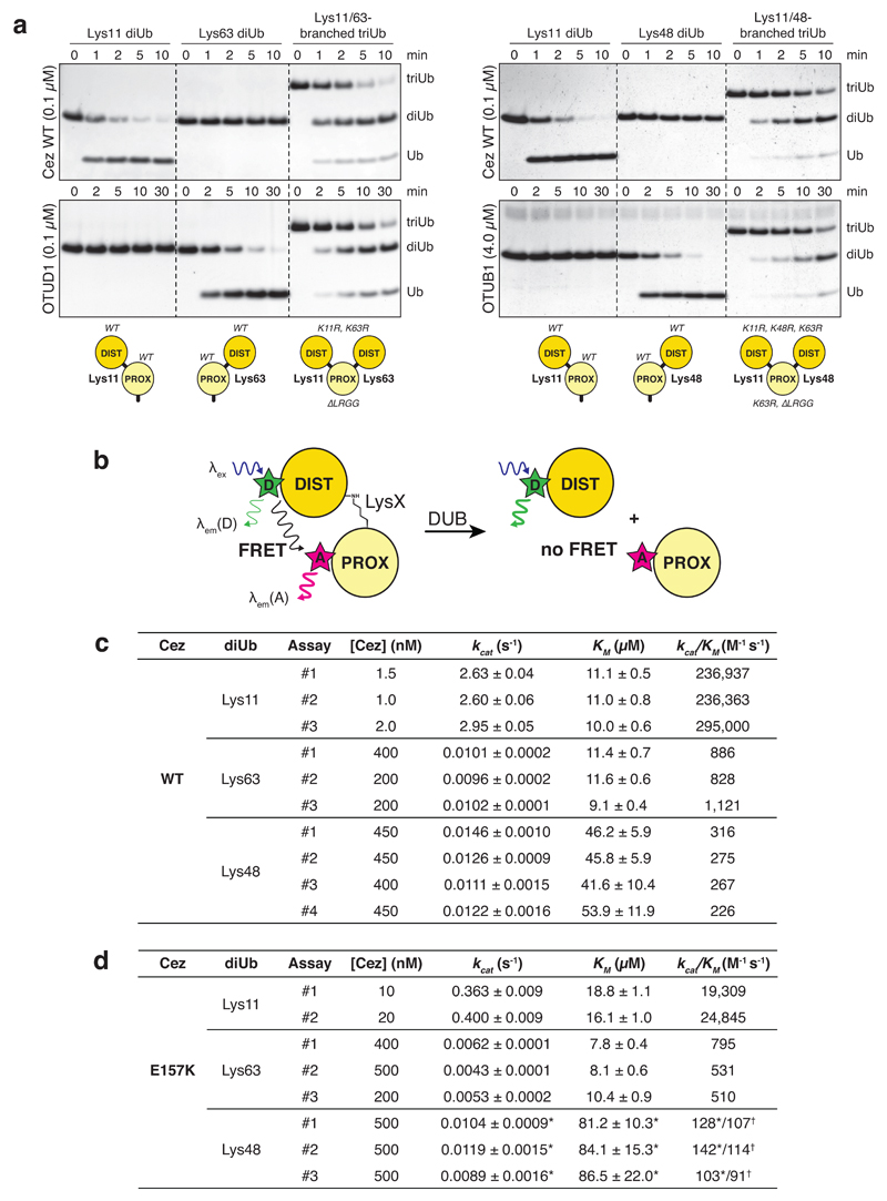
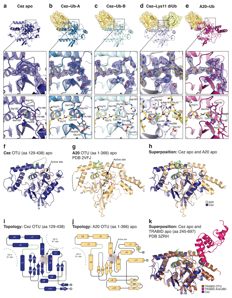
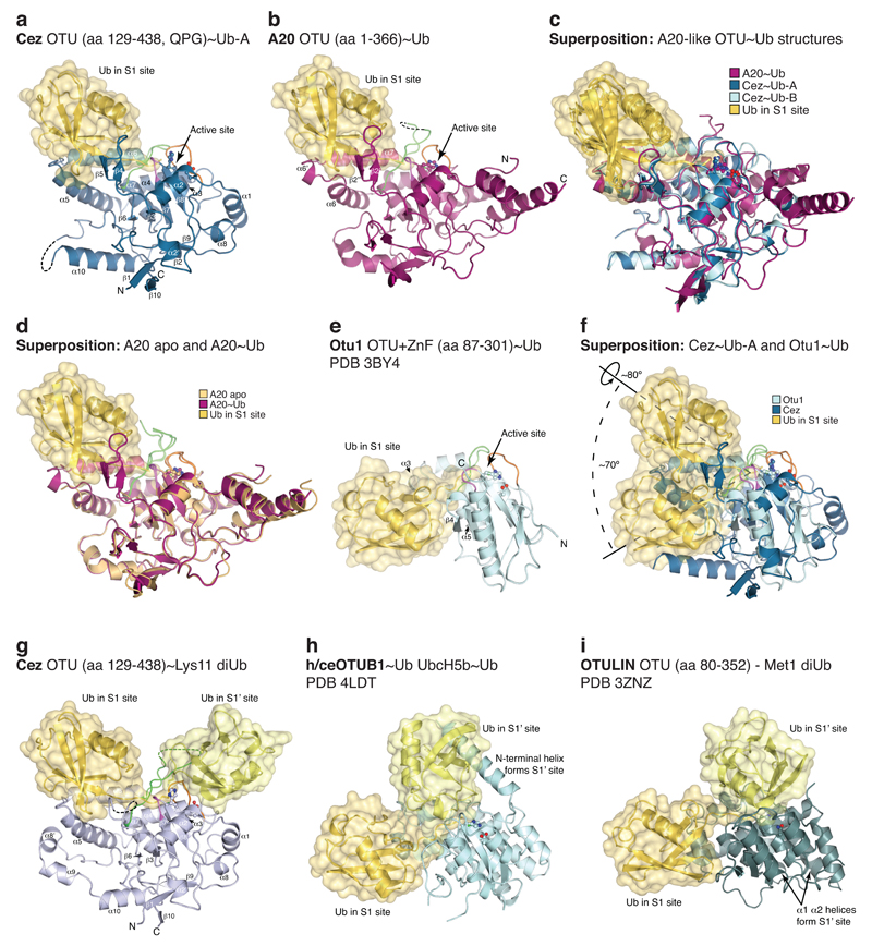
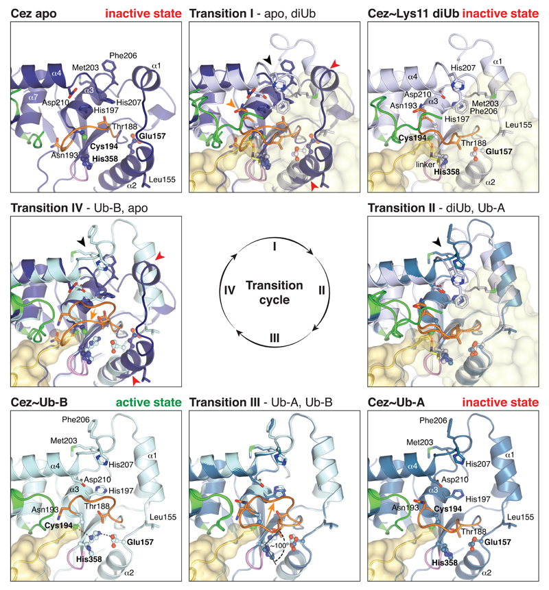
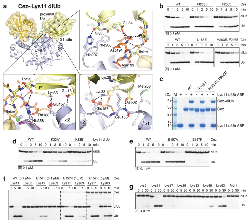
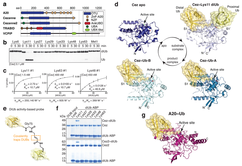
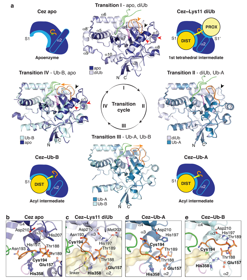
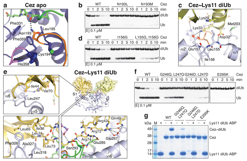
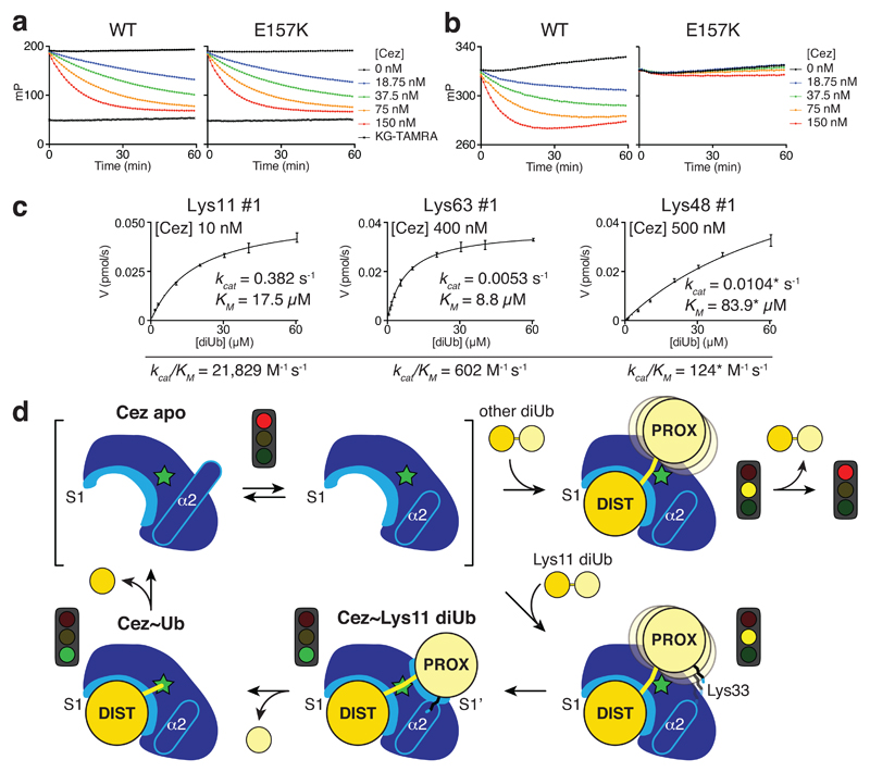
The post-translational modification of proteins with polyubiquitin regulates virtually all aspects of cell biology. Eight distinct chain linkage types co-exist in polyubiquitin and are independently regulated in cells. This 'ubiquitin code' determines the fate of the modified protein. Deubiquitinating enzymes of the ovarian tumour (OTU) family regulate cellular signalling by targeting distinct linkage types within polyubiquitin, and understanding their mechanisms of linkage specificity gives fundamental insights into the ubiquitin system. Here we reveal how the deubiquitinase Cezanne (also known as OTUD7B) specifically targets Lys11-linked polyubiquitin. Crystal structures of Cezanne alone and in complex with monoubiquitin and Lys11-linked diubiquitin, in combination with hydrogen-deuterium exchange mass spectrometry, enable us to reconstruct the enzymatic cycle in great detail. An intricate mechanism of ubiquitin-assisted conformational changes activates the enzyme, and while all chain types interact with the enzymatic S1 site, only Lys11-linked chains can bind productively across the active site and stimulate catalytic turnover. Our work highlights the plasticity of deubiquitinases and indicates that new conformational states can occur when a true substrate, such as diubiquitin, is bound at the active site.


Conflict of interest statement

Statement D.K. and H.O. are part of the DUB Alliance that includes Cancer Research Technology and FORMA Therapeutics. H.O. and F.E. are co-founders and shareholders of UbiQ Bio BV.

Figures

Extended Data Figure 1
Extended Data Figure 1. Analysis of branched triUb substrates and FRET-based diUb cleavage kinetics
a, Branched triUb molecules with different topology were generated as shown in the schematic (bottom). Lys11 diUb, Lys63 diUb and branched Lys11/63 triUb (left panel) were treated with Cez WT (top) and OTUD1 (residues 287-481, bottom), a Lys63-specific enzyme. Both DUBs cleaved their preferred diUb substrate as well as one linkage of the branched triUb molecule. Lys11 diUb, Lys48 diUb and branched Lys11/48 triUb (right panel) were incubated with Cez WT (top) and OTUB1 (full-length, bottom), a Lys48-specific OTU DUB. Again, both enzymes showed similar activities towards their preferred linkage type in a diUb substrate and a branched triUb molecule. This shows that Cezanne can cleave Lys11 linkages in the context of Lys11/Lys63- and Lys11/Lys48-branched chains. For gel source data, see Supplementary Figure 1. b, Schematic of FRET-based diUb cleavage assays to derive DUB kinetics. Distal and proximal Ub moieties were modified with a donor (D) and acceptor (A) fluorophore, respectively. Upon DUB treatment, the native isopeptide bond is cleaved and the FRET signal is lost. The increase in donor intensity is measured to follow the reaction. c, Kinetic parameters for all independently performed experiments of Lys11, Lys63 and Lys48 diUb cleavage by Cez WT. Values are in good agreement with previously published parameters derived from gel-based studies. d, Summary of kinetic parameters for Lys11-, Lys63- and Lys48-linked diUb cleavage by Cez E157K. The determined KM values for Lys48 diUb lie above the highest tested substrate concentration, so kinetic parameters marked by an asterisk (*) were calculated from experiments where substrate saturation could not be achieved due to technical limitations. Catalytic efficiencies (kcat/KM) for this substrate were also derived from a linear fit of the lower concentration range (0-20 μM, linear part of the graph). These values are marked by a cross (). The similarity of catalytic efficiencies calculated in two different ways indicate that the kinetic parameters marked by an asterisk (*) are good estimates. See Supplementary Figure 2 for all corresponding graphs of initial rates.
Extended Data Figure 2
Extended Data Figure 2. Crystal structures determined in this study and comparison of A20-like OTU apo structures
a-e, Active site regions Cez apo (a), Cez~Ub-A (b), Cez~Ub-B (c), Cez~Lys11 diUb (d), and A20~Ub (e). 2|FO| – |FC| electron density maps contoured at 1σ (blue) cover catalytic residues, the Cys-loop and chemical linkers in the complex structures. Hydrogen bonds between the oxyanion hole and the Lys11 diUb ABP linker carbonyl are indicated in d, and the sp3-hybridised carbon atom that is linked to the oxyanion in a native first tetrahedral intermediate is highlighted (green arrow). f, g, Cezanne OTU (as in Fig. 1d) and A20 OTU (PDB 2VFJ, 22) apo structures with labelled secondary structure elements. Catalytic residues are shown in ball-and-stick representation. Three loops surrounding the active site are coloured (Cys-loop, orange; V-loop, green; His-loop, purple). h, Superposition of f and g showing structural similarities and differences between Cezanne and A20. i, j, Topology diagrams of f and g. The catalytic centre is indicated (red stars) and Ub-binding sites are highlighted. A20 contains two additional N-terminal and one additional C-terminal helices compared to Cezanne. The β1-β10 sheet in Cezanne corresponds to the A20 β7-β8 sheet. This explains why sequence-based alignments are challenging. k, Superposition of Cez apo (f) and TRABID AnkUBD (pink) and OTU (brown) domains (residues 245-697, PDB 3ZRH, 10).
Extended Data Figure 3
Extended Data Figure 3. Comparison of Ub and diUb complexes within the OTU family
Ub moieties are shown in cartoon representation under transparent surfaces in shades of yellow. Secondary structure elements involved in Ub binding are labelled, and active site loops are coloured as in Extended Data Fig. 2f. a, Cez~Ub-A complex as in Fig. 1d. b, A20~Ub complex as in Fig. 1g. c, Superposition of Ub complexes reveals a conserved S1 Ub-binding mode in A20-like OTU DUBs. d, Superposition of A20 apo (Extended Data Fig. 2g) and A20~Ub (b). No large conformational changes occur upon Ub binding. However, two unstructured loops in A20 apo are stabilised by Ub, forming helix α6’ and the β2’-β2” sheet (compare Extended Data Fig. 2j). e, The structure of the yeast Otu1~Ub complex (PDB 3BY4,42) is representative for the OTUD subfamily of OTU DUBs. The Ub moiety in the S1 site is mainly bound by the short helix α3. f, The superposition of Cez~Ub (a) and Otu1 (e) reveals substantially different S1 sites between A20-like and OTUD subfamilies. Rotations around the roll axis of Ub (~80°) and the active site (~70°) would be required to align both Ub moieties. g-i, Structures of OTU domains in identical orientation bound to their respective diUb substrate. The binding mode of proximal and distal Ub differs dramatically between the here determined Cez~Lys11 diUb complex (g, as shown in Fig. 1d), the h/ceOTUB1~Ub UbcH5b~Ub structure (PDB 4LDT, , UbcH5b molecule is not shown), which resembles an OTUB1 Lys48 diUb complex (h), and OTULIN bound to Met1-linked diUb (PDB 3ZNZ, 3) (i).
Extended Data Figure 4
Extended Data Figure 4. Conformational changes in the catalytic centre
Cezanne structures according to Fig. 2a are shown in the corners, and Transitions I-IV are overlays of neighbouring structures. Side chains of catalytic residues and other selected residues are highlighted. Loops are coloured as in Extended Data Fig. 2f. Cez apo shows a catalytically incompetent state. His358 and Glu157 are in flipped-out conformations. Transition I features structural rearrangements of the Cys-loop (orange arrow), helices α1 and α2 (red arrows) and the S1’-loop (black arrow). Cez~Lys11 diUb also features an inactive state; His358 remains flipped-out, which is caused by the Cys-loop residue Thr188 that is pushed into the active site by the proximal Ub. In Transition II, another S1’-loop movement relocates S1’ site residues (black arrow). A similar inactive state is present in Cez~Ub-A, and Thr188 still resides in the active site. The absence of the proximal Ub allows the Cys-loop and Thr188 to move in Transition III (orange arrow), allowing a ~100° rotation of His358. Hence, Cez~Ub-B contains an aligned catalytic centre. Hydrogen bonds are indicated. In Transition IV, large conformational changes in various parts of the OTU domain regenerate the autoinhibited apoenzyme.
Extended Data Figure 5
Extended Data Figure 5. Ub binding to Cezanne and mutational analysis of residues involved in catalysis and conformational dynamics
a, NMR analysis of Ub binding to Cez WT and the covalent Cez~Ub complex. 1H-15N BEST-TROSY spectra of 50 μM 15N-labelled Ub alone (black) and in the presence of 130 μM unlabelled Cez WT (red, left) or unlabelled Cez~Ub (red, right). Strong chemical shift perturbations upon addition of Cez WT indicate binding to Ub. In contrast, no chemical shifts were detected with Cez~Ub, suggesting that all changes with Cez WT can be attributed to the S1 site (this site is occupied by unlabelled Ub in Cez~Ub). More importantly, this also indicates that a functional S1’ site is not present in the Cez~Ub complex. b, Fluorescence polarisation (FP) experiment assessing the binding of FlAsH-tagged Ub to catalytically inactive Cez (C194A), Cez WT, an S1 site mutant (E295K, see below) and the Cez~Ub complex. c, Lys11 diUb cleavage assays of catalytic Cys194 and His358 mutants. d, e, Ub-KG* cleavage by catalytic Cys194 and His358 mutants (d), as well as Asn193 and helix α2 mutants that modulate the overall dynamics of Cezanne (e). This assay follows fluorescent dye release in the reaction; the fact that Cez H358A is inactive indicates an important role in the deprotonation of the catalytic Cys at the start of the reaction (i.e. the catalytic centre transiently adopts an active state) and/or a role in resolving the first tetrahedral intermediate. In case His358 was not required for either, we would expect a single turnover of the reaction, which would stop at the thioester intermediate. The release of KG-TAMRA would still occur, but was not detected even at an enzyme concentration of 150 nM (the substrate concentration in all assays was 150 nM). The FP signal also did not increase, suggesting that no covalent first tetrahedral intermediate was formed due to impaired dye release. Hence, the data suggests a role for His358 at least in the initial Cys deprotonation in addition to the last reaction step. f, FP binding assay of Asn193 and helix α2 mutants compared to constructs used in b. g, h, Hydrolysis of Lys11-linked diUb (g) and Ub-KG* (h) by Cez H197A and D210A. i, DUB assay with Cezanne variants (extended incubation at room temperature, RT). j, Lys11 diUb cleavage assay with His197 variants. k, FP binding assay as in b testing His197 variants. l, Mutation of corresponding residues in A20 (A20 His256 corresponds to Cez His358, and A20 His106 to Cez His197) have similar effects on Lys48 diUb hydrolysis. All DUB assays are representative of at least two independent experiments for every construct. Ub-KG* cleavage experiments and FP binding assays were replicated at least twice for each variant with consistent results. FP measurements were performed in triplicate. Error bars represent standard deviation from the mean. mP, millipolarisation unit. For gel source data, see Supplementary Figure 1.
Extended Data Figure 6
Extended Data Figure 6. HDX-MS analysis of the Cez apo state
a, HDX-MS experiment showing the conformational dynamics of Cez WT. The relative fractional deuterium uptake is shown for four time points (0.3-300 s). Protein sequence and secondary structure elements of Cez apo (dark grey) and Cez~diUb (light grey) are aligned. Residues of the catalytic centre are indicated by stars. b, Cez apo structure coloured based upon the relative fractional deuterium uptake of Cez WT at 0.3 s, 3 s, 30 s and 300 s. The region spanning helices α1 and α2 shows a particularly high deuterium uptake, suggesting conformational flexibility in this region in solution. c, The H/D exchange of the α2-helix destabilising mutant Cez L155G/I156G compared to Cez WT. Cez apo structure coloured based upon the difference in deuterium uptake (L155G/I156G-WT) at 0.3 s, 3 s, 30 s and 300 s (heat maps are shown in Supplementary Figure 3). The data suggest that helix α2 is destabilised, as regions structurally adjacent to the mutation site (black arrow) show an increased deuterium uptake as compared to Cez WT. Peptides containing the mutations could not be analysed due to the different sequences, and are therefore coloured in grey. Notably, most differences are stronger at shorter time points, indicating increased dynamics within this time frame (0.3-30 s). At the latest time point (300 s), differences are not as pronounced, suggesting that Cez WT undergoes the same structural rearrangements at a slower speed. Importantly, the data also confirms that overall folding of the mutant was not affected by the two Gly residues introduced in helix α2.
Extended Data Figure 7
Extended Data Figure 7. HDX-MS analysis of Transitions I, II and IV
a, HDX-MS experiments were performed with Cez apo, Cez~Lys11 diUb and Cez~Ub. Heat maps show differences in deuterium uptake between two states in each case: Transition I (diUb-apo), Transition II (Ub-diUb) and Transition IV (apo-Ub). Hence, Cezanne regions that are stabilised or more protected upon Lys11 diUb binding (Transition I), or more flexible or exposed upon the stepwise release of the proximal Ub (Transition II) and the distal Ub (Transition IV) are highlighted. The S1 site predominantly consists of helices α5 and α6 (i.e. helical content with very low deuterium uptake in any state), and is not as easily detected as the S1’ site that features various loops and the dynamic helix α2. Cezanne sequence and secondary structure schematics are shown as in Extended Data Fig. 6a. b, Cez~Lys11 diUb structure (shown without Lys11 diUb) coloured based upon Transition I deuterium uptake at 30 s. c, Transition II deuterium uptake at 30 s plotted onto Cez~Ub-B (shown without Ub). d, Cez apo coloured based upon Transition IV deuterium uptake at 30 s.
Extended Data Figure 8
Extended Data Figure 8. Mutational analysis of the S1 Ub-binding site
a, Thermal shift assay of Cez WT and C194A. In the presence of Ub, the melting temperature (Tm) of Cezanne increases. Data were recorded in triplicate and in two independent experiments. b, Ub-KG* hydrolysis by S1 site mutants. c, d, FP-based affinity measurement using N-terminally FlAsH-tagged Ub. Dissociation constants (Kd values) for Cez WT (c), C194A and C194S (d) are shown. Data are representative of at least two independent experiments per construct. e, Pull-down assay with His-tagged Cez constructs (catalytically inactive C194A, S1 site mutant C194A/E295K or WT) and different Ub and diUb variants. MonoUb requires an intact C-terminus to bind to Cez C194A. To prevent unspecific binding of differently linked diUb molecules with their proximal Ub to the S1 site, the C-terminus was removed (ΔLRGG). Variants marked by an asterisk (*) were assembled by using K11R, S20C and K63R mutations in the distal Ub, as well as K63R (only for Lys11 diUb) and ΔLRGG in the proximal Ub moiety. Pull-down and input samples were analysed by SDS-PAGE and silver staining. The pull-down assay was performed in two independent experiments. For gel source data, see Supplementary Figure 1.
Extended Data Figure 9
Extended Data Figure 9. Biochemical analysis of S1’ site mutations
a, The interface between Cezanne and the proximal Ub in the Cez~Lys11 diUb complex. An unusual surface of Ub comprising Glu16, Asp32, Lys33 and Glu34 is contacted by the S1’ site (Leu155, Glu157, Met203, Phe206 and His207). b, c, Lys11 diUb cleavage (b) and Lys11 diUb ABP reactivity (c) assays with S1’ site mutants. d, DUB assays with Cez WT and Ub variants. Lys11 diUb substrates were assembled to specifically mutate the proximal Ub by using K11R, K63R mutations in the distal, and K63R, ΔLRGG in the proximal Ub moiety. No further mutations were introduced in WT*, while K33A* and K33E* variants additionally contained respective mutations in their proximal Ub only. e, Lys11 diUb cleavage assay with Glu157 variants. f, g, Gel-based specificity analysis of Cez E157K. The mutant shows a reduced activity towards Lys11-linked diUb and therefore specificity compared to Cez WT (compare Fig. 1b). Assays with each variant were performed at least twice with consistent results. For gel source data, see Supplementary Figure 1.
Figure 1
Figure 1. Cezanne biochemistry and structures
a, Domain architecture of A20-like OTU DUBs. b, Specificity analysis of the Cezanne OTU domain (residues 129-438). This experiment was performed three times. c, Representative graphs of initial rates and kinetic parameters for Lys11, Lys63 and Lys48 diUb hydrolysis by Cezanne. Assays were performed in triplicate and in at least three independent experiments (Extended Data Fig. 1c). Error bars represent standard deviation from the mean. d, Cezanne (residues 129-438) structures determined in this study: Cez apo, Cez~Lys11 diUb and Cez~Ub (‘QPG’) (see Methods). Both Cez~Ub complexes in the asymmetric unit are depicted. The OTU domain is shown as cartoon with active site residues highlighted, and Ub moieties are shown under transparent surfaces. e, Schematic of diUb ABPs. f, Probe assay of Cezanne (residues 129-438, top) and Cezanne2 (residues 1-462, bottom) with differently linked diUb ABPs. Experiments were replicated twice. g, Crystal structure of A20~Ub (residues 1-366, see Methods). For gel source data, see Supplementary Figure 1.
Figure 2
Figure 2. Conformational changes in the Cezanne catalytic cycle
a, Schematic cartoons of the determined structures (Fig. 1d) highlight four catalytic states of the reaction cycle (green star, active site; orange line, Cys-loop). In between, superpositions of the OTU domain show Transitions I-IV. Loops are coloured orange (Cys-loop), green (V-loop) and purple (His-loop). Transition I (diUb substrate binding) is characterised by conformational changes around the catalytic centre, including the Cys-loop (orange arrow), α1 and α2 helices (red arrows) and the S1’-loop (black arrow). In Transition II (proximal Ub release), a second S1’-loop rearrangement relocates S1’ site residues (black arrow). Transition III features a Cys-loop movement. Several structural changes regenerate Cez apo in Transition IV. Also see Supplementary Video 1. b-e, Active site close-up of the four states. Selected residues are shown as sticks. Hydrogen bond networks of His197 and the catalytic centre are indicated. Also see Extended Data Fig. 4 and Supplementary Video 2.
Figure 3
Figure 3. Mutational analysis of Cezanne dynamics
a, In Cez apo, Asn193 of the Cys-loop (orange) blocks the distal Ub-binding channel and is positioned above a hydrophobic pocket. b, DUB assay of Asn193 mutants that ‘lock’ the enzyme in the apo state. c, The α1-α2 linker in Cez~diUb contains residues Leu155 and Ile156 adjacent to Glu157. d, DUB assay of mutants with a destabilised α2-helix (see Extended Data Fig. 6c). e, Cezanne’s S1 site contacts the Ile44 patch (top left), the Ile36 patch (bottom left) and the C-terminal tail of the distal Ub (bottom right). f, DUB assay of S1 site mutants. g, Lys11 diUb ABP probe assay of S1 site mutants.All assays were performed at least twice with consistent results. For gel source data, see Supplementary Figure 1.
Figure 4
Figure 4. Basis of Lys11 specificity and model of Cezanne mechanism
a, b, FP cleavage assays comparing Cez WT and E157K using Ub-KG* (a) and FlAsH-tagged Lys11-linked diUb (b). FP measurements were performed in triplicate in at least two independent experiments. c, Summary of Cez E157K diUb cleavage kinetics. Compared to Cez WT (Fig. 1c), this mutant is impaired in cleaving Lys11 linkages. Assays were performed in triplicate and in at least two independent experiments. Values marked by an asterisk (*) suffer from technical limitations (for more detail, see Extended Data Fig. 1d). Error bars represent standard deviation from the mean. d, Model of Cezanne mechanism. The apoenzyme is autoinhibited yet dynamic, and recruits a substrate with its accessible S1 site. Only Lys11-linked diUb can interact with the formed S1’ site specifically, involving an activating interaction between Cezanne and Ub Lys33. After cleavage, the S1’ site is lost and the proximal moiety expelled. Subsequent hydrolysis and distal Ub release recreates the Cezanne apo state.

References

    1. Komander D, Rape M. The ubiquitin code. Annu Rev Biochem. 2012;81:203–229. - PubMed
    1. Mevissen TET, et al. OTU Deubiquitinases Reveal Mechanisms of Linkage Specificity and Enable Ubiquitin Chain Restriction Analysis. Cell. 2013;154:169–184. - PMC - PubMed
    1. Keusekotten K, et al. OTULIN antagonizes LUBAC signaling by specifically hydrolyzing Met1-linked polyubiquitin. Cell. 2013;153:1312–1326. - PMC - PubMed
    1. Wiener R, Zhang X, Wang T, Wolberger C. The mechanism of OTUB1-mediated inhibition of ubiquitination. Nature. 2012;483:618–622. - PMC - PubMed
    1. Juang Y-C, et al. OTUB1 Co-opts Lys48-Linked Ubiquitin Recognition to Suppress E2 Enzyme Function. Mol Cell. 2012;45:384–397. - PMC - PubMed

MeSH terms