. 2016 Oct 11;17(3):735-747.
doi: 10.1016/j.celrep.2016.09.033.

Astroglial-Mediated Remodeling of the Interhemispheric Midline Is Required for the Formation of the Corpus Callosum

Affiliations

Astroglial-Mediated Remodeling of the Interhemispheric Midline Is Required for the Formation of the Corpus Callosum

Ilan Gobius et al. Cell Rep. .

Abstract

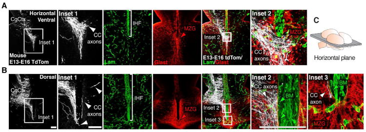
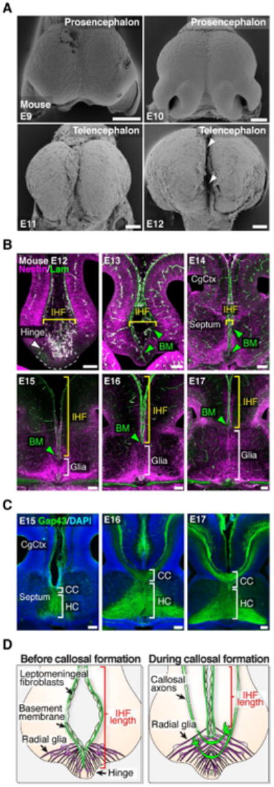
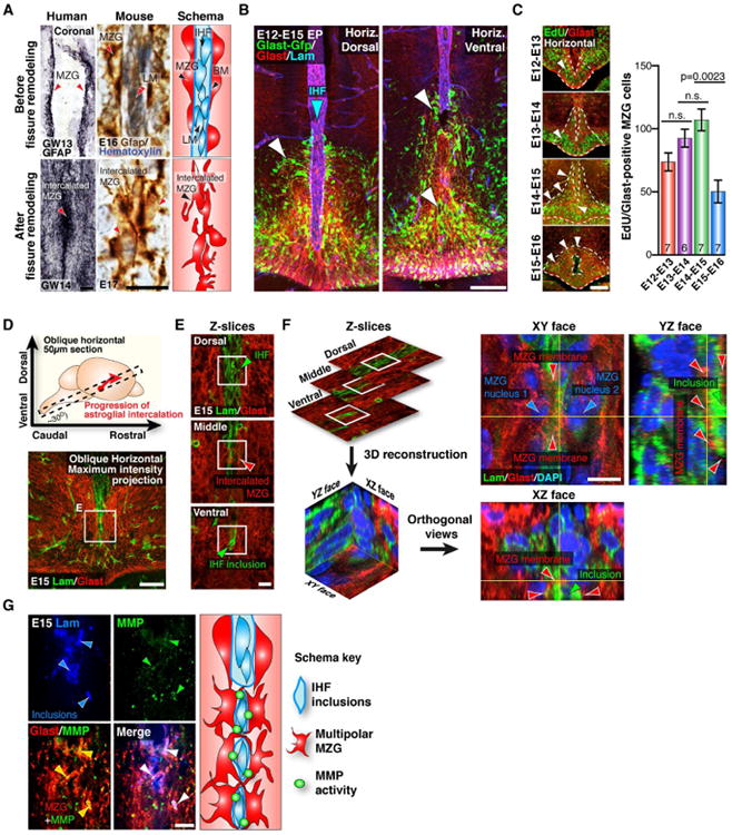
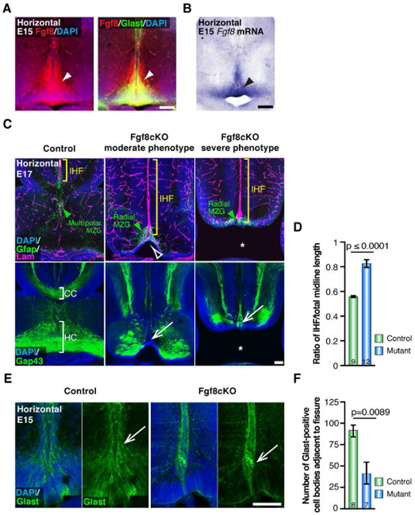
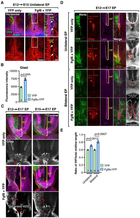
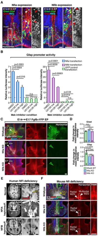
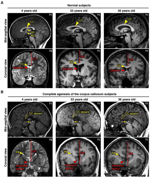
The corpus callosum is the major axon tract that connects and integrates neural activity between the two cerebral hemispheres. Although ∼1:4,000 children are born with developmental absence of the corpus callosum, the primary etiology of this condition remains unknown. Here, we demonstrate that midline crossing of callosal axons is dependent upon the prior remodeling and degradation of the intervening interhemispheric fissure. This remodeling event is initiated by astroglia on either side of the interhemispheric fissure, which intercalate with one another and degrade the intervening leptomeninges. Callosal axons then preferentially extend over these specialized astroglial cells to cross the midline. A key regulatory step in interhemispheric remodeling is the differentiation of these astroglia from radial glia, which is initiated by Fgf8 signaling to downstream Nfi transcription factors. Crucially, our findings from human neuroimaging studies reveal that developmental defects in interhemispheric remodeling are likely to be a primary etiology underlying human callosal agenesis.

Keywords: Fgf8; Nfia; Nfib; astrocyte; callosal agenesis; interhemispheric fissure.


Figures

Figure 1
Figure 1. Pioneer callosal axons do not cross through the IHF but rather use the MZG as a growth substrate to cross the midline
(A-B) Electroporation of the mouse cingulate cortex (CgCtx) at E13 with a myristoylated tdTomato reporter plasmid (white) labels pioneering callosal axons at E16, visualized in relation to Glast-positive MZG cells (red) and the Laminin-positive interhemispheric fissure (IHF; green) in ventral (A) and dorsal (B) horizontal sections. Callosal axons (white arrowheads) project around the basement membrane (BM) of the IHF (green arrowheads) and across the midline over MZG cells (red arrowheads). (C) Schematic showing the orientation of the horizontal plane shown in A and B. CC, corpus callosum. Scale bars, 100μm.
Figure 2
Figure 2. Developmental remodeling of the IHF is associated with callosal tract formation in mice
(A) Frontal view scanning electron microscopy photomicrographs show the development of the mouse IHF (arrowheads). (B) Fluorescence immunohistochemistry for Nestin-positive radial glia (magenta) and pan-Laminin-positive leptomeninges and basement membrane (BM; green) from E12 to E17. Brackets indicate the spatial extent of radial glia and IHF. Green arrowheads denote the BM. (C) Fluorescence immunohistochemistry for Gap43 labeling corpus callosum (CC) and hippocampal commissure (HC) axons from E15 to E17. (D) Schema of the midline remodeling events occurring before and during callosal tract formation. CgCtx, cingulate cortex. Scale bars, 100 μm. See also Figure S1.
Figure 3
Figure 3. The transition of radial MZG into multipolar astroglia initiates IHF remodeling and callosal tract formation
(A) Embryonic human and mouse brain sections immunostained for Gfap (black and brown, respectively), counterstained with hematoxylin (blue) in mouse sections. Gfap-positive MZG cells (arrowheads) are schematically represented far-right panel. (B) In utero electroporation (EP) of the mouse telencephalic hinge with piggyBac Glast transposase/GFP reporter plasmids (green), colabeled for Glast (red) and Laminin (blue) imunohistochemistry in dorsal and ventral sections. GFP labeling shows both MZG cells (white arrowheads) undergoing somal translocation towards the IHF (blue arrowhead) and MZG cells undergoing intercalation. (C) Glast-positive mouse MZG cells (red) were birthdated with the thymidine analogue EdU (green) every 24 hours, from E12 to E15, quantified on the right. Arrowheads show distribution of MZG progenitors within the telencephalic hinge niche (dotted lines). (D) Schema of the oblique horizontal sectioning plane (top) and confocal maximum intensity projection of an E15 oblique horizontal section (bottom) immunostained with Laminin (green) and Glast (red). (E) Optical sectioning of the region shown in (D). (F) Reconstruction of z-planes in (E) to resolve the XY, XZ and YZ orthogonal views of the MZG-IHF interface, showing MZG projections (red arrowheads) partitioning laminin-positive inclusions of the IHF (green arrowheads). (G) Pan-matrix metalloproteinase activity labeled with MMPSense (green) is associated with Glast-positive glial membranes adjacent to Laminin-positive IHF inclusions (blue), with schema (right). BM= basement membrane, LM = leptomeninges. Data are represented as means ± SEM (n-values within bars). Scale bars, 50μm (A, left), 5μm (A, right), 100 μm (B, C, and D), 10μm (E, F and G). See also Figure S2.
Figure 4
Figure 4. Fgf8 signaling is required for MZG development and interhemispheric remodeling in mice
(A) Fluorescence immunohistochemistry shows expression of Fgf8 (red) in Glast-positive radial MZG (green) at E15 along the interhemispheric fissure (IHF; arrowheads). (B) In situ hybridization for Fgf8 mRNA. (C) Immunohistochemistry for Gfap (green), Laminin (magenta) and Gap43 (green) in E17 Fgf8 conditional knockout (cKO) mice and littermate controls. Note thinning of the MZG progenitor niche in moderate phenotype mutants (open arrowhead) versus complete absence of septum in severe mutants (asterisk). Green arrowheads indicate Gfap-positive MZG, yellow brackets indicate the IHF, and white arrows indicate complete agenesis of the corpus callosum and hippocampal commissure. (D) Quantification of IHF length in E17 Fgf8 cKO and control brains. (E) Immunohistochemistry for Glast (green) in E15 Fgf8cKO mutants and littermate controls, note reduction in Glast-positive MZG adjacent to the fissure pial surface in Fgf8cKO mutants (arrows). (F) Quantification of the number of Glast-positive cell bodies adjacent to the IHF in E15 Fgf8 cKO mice and littermate control brains. Data are represented as means ± SEM (n-values within bars). Scale bars, 100 μm. See also Figure S3.
Figure 5
Figure 5. Fgf8 signaling promotes astroglial maturation of the MZG in mice
(A) Unilateral E12→E15 in utero electroporation (EP) of YFP alone or coelectroporation of Fgf8 + YFP plasmids followed by fluorescence immunohistochemistry for YFP (green) and Glast (red). Arrowheads indicate excess Glast-positive cell bodies along the pial surface of the Fgf8-electroporated hemisphere. (B) Quantification of Glast fluorescence intensity in YFP versus Fgf8+YFP electroporated hemispheres. (C) E12→E17 and E15→E17 unilateral EP of YFP alone or coelectroporation of Fgf8 + YFP plasmids followed by fluorescence immunohistochemistry for YFP (green), Gfap (red and white) and Gap43 (magenta) in coronal sections. Double red arrowheads indicate precocious Gfap expression following Fgf8 overexpression, brackets indicate the length of the IHF and the presence/absence of the corpus callosum (CC) in each condition, and single red arrowheads indicate the distribution of the MZG. (D) Unilateral or bilateral E12→E17 EP of Fgf8 + YFP followed by fluorescence immunohistochemistry for YFP, Gfap, Laminin and Gap43 in horizontal sections. Brackets indicate the IHF length in each condition, and arrowheads indicate the distribution of MZG cells. Note dysgenesis or agenesis of the CC in Fgf8 EP conditions. (E) Quantification of IHF length in unilateral and bilateral E12→E17 Fgf8 + YFP and YFP control electroporated brains. Data are represented as means ± SEM (n-values within bars). Scale bars, 100 μm (A, left; C and D), 20μm (20μm). See also Figure S4.
Figure 6
Figure 6. Nfia and Nfib are downstream transcriptional effectors of Fgf8 signaling required for IHF remodeling and corpus callosum formation
(A) E12→E15 in utero electroporation (EP) of the mouse telencephalic hinge with piggyBac Glast transposase/GFP (green) reporter plasmids, colabeled with Glast (red) and either Nfia or Nfib (blue/white) demonstrates Nfia and Nfib are both expressed by radial MZG (arrowheads). (B) Luciferase activity of U251 cells cotransfected with a Gfap promoter luciferase construct and either Nfia, Nfib or GFP control expression constructs, following addition of the Mek inhibitor U0126 (low = 5 μM; high = 20 μM), performed in triplicate. Data are represented as mean luciferase intensity ± SD, normalized to the GFP DMSO control (dotted line) and are representative of at least 2 independent experiments. Significant differences were determined with a Student's t-test. (C) Unilateral E14→E17 EP of Fgf8 + YFP plasmids into Nfia and Nfib knockout (Nfi KO) mice and their wildtype littermates, followed by fluorescence immunohistochemistry for Gfap (green) and Glast (red). Note increase in Gfap expression in wildtypes (closed green arrowhead) and absence of Gfap expression in Nfi KO mice (open green arrowheads). (D) Quantification of the fold change in Gfap and Glast fluorescence intensity. Data are represented as means ± SEM (n-values within bars, significant differences determined with a non-parametric Mann-Whitney test). (E) Coronal and axial structural T1- and T2-weighted MRI images of a normal human brain compared with NFIA- and NFIB-haploinsufficient individuals. Brackets indicate interhemispheric fissure (IHF) length and red arrowheads indicate the separated septum in NFI haploinsufficient brains. (F) Fluorescence immunohistochemistry for Glast (red), Laminin (green) and Gap43 (white) in Nfia and Nfib KO embryos and wildtype littermates at E16. Brackets indicate IHF length and white arrowheads indicate the distribution of radial versus multipolar MZG cells. Yellow arrowhead indicates the callosal (CC) and hippocampal commissural (HC) tract in wildtypes. Scale bars, 50μm (A), 100 μm (C and F) and 1 cm (E). See also Figure S5.
Figure 7
Figure 7. Characterization of human IHF phenotypes associated with agenesis of the corpus callosum based on structural MRI studies
(A and B) Comparison of T1- or T2-weighted structural MRI images from 3 individuals previously diagnosed with isolated complete agenesis of the corpus callosum and age-matched controls. Note abnormal retention of the IHF and accumulation of callosal axons into Probst bundles (PB) either side of the IHF in individuals with callosal agenesis. Arrowheads indicate the morphology of the septum (red arrowheads) and corpus callosum (CC; yellow arrowheads). AC = anterior commissure, HC = hippocampal commissure. See also Table S1.

References

    1. Bagri A, Marin O, Plump AS, Mak J, Pleasure SJ, Rubenstein JL, Tessier-Lavigne M. Slit proteins prevent midline crossing and determine the dorsoventral position of major axonal pathways in the mammalian forebrain. Neuron. 2002;33:233–248. - PubMed
    1. Barton JS, O'Loughlin J, Howell RT, L'E Orme R. Developmental delay and dysmorphic features associated with a previously undescribed deletion on chromosome 1. Journal of medical genetics. 1995;32:636–637. - PMC - PubMed
    1. Bremer C, Tung CH, Weissleder R. In vivo molecular target assessment of matrix metalloproteinase inhibition. Nature medicine. 2001;7:743–748. - PubMed
    1. Chen CP, Su YN, Chen YY, Chern SR, Liu YP, Wu PC, Lee CC, Chen YT, Wang W. Chromosome 1p32-p31 deletion syndrome: prenatal diagnosis by array comparative genomic hybridization using uncultured amniocytes and association with NFIA haploinsufficiency, ventriculomegaly, corpus callosum hypogenesis, abnormal external genitalia, and intrauterine growth restriction. Taiwanese journal of obstetrics & gynecology. 2011;50:345–352. - PubMed
    1. Chen F, LoTurco J. A method for stable transgenesis of radial glia lineage in rat neocortex by piggyBac mediated transposition. J Neurosci Methods. 2012;207:172–180. - PMC - PubMed

Publication types

Substances