. 2020 Jun 15;37(12):1452-1462.
doi: 10.1089/neu.2016.4644. Epub 2016 Nov 2.

Cerium Oxide Nanoparticles Improve Outcome after In Vitro and In Vivo Mild Traumatic Brain Injury

Affiliations

Cerium Oxide Nanoparticles Improve Outcome after In Vitro and In Vivo Mild Traumatic Brain Injury

Zachary S Bailey et al. J Neurotrauma. .

Abstract

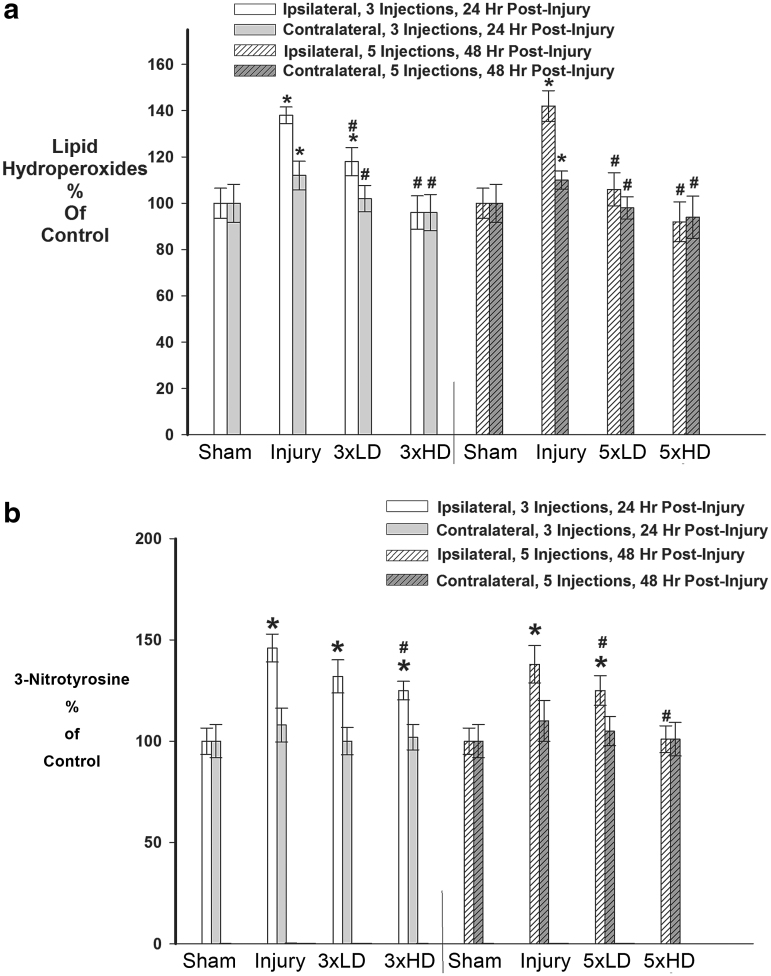
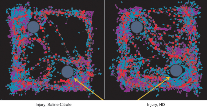
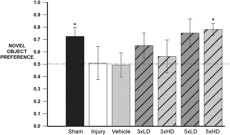
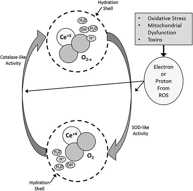
Mild traumatic brain injury results in aberrant free radical generation, which is associated with oxidative stress, secondary injury signaling cascades, mitochondrial dysfunction, and poor functional outcome. Pharmacological targeting of free radicals with antioxidants has been examined as an approach to treatment, but has met with limited success in clinical trials. Conventional antioxidants that are currently available scavenge a single free radical before they are destroyed in the process. Here, we report for the first time that a novel regenerative cerium oxide nanoparticle antioxidant reduces neuronal death and calcium dysregulation after in vitro trauma. Further, using an in vivo model of mild lateral fluid percussion brain injury in the rat, we report that cerium oxide nanoparticles also preserve endogenous antioxidant systems, decrease macromolecular free radical damage, and improve cognitive function. Taken together, our results demonstrate that cerium oxide nanoparticles are a novel nanopharmaceutical with potential for mitigating neuropathological effects of mild traumatic brain injury and modifying the course of recovery.

Keywords: cerium oxide; nanoparticles; oxidative stress; traumatic brain injury.


Conflict of interest statement

No competing financial interests exist.

Figures

FIG. 1.
FIG. 1.
Cerium oxide nanoparticles. Representative transmission electron microscopy (TEM) image of the nanoparticle solution as delivered, in saline-citrate buffer (average particle size 10 nm). Note lack of agglomeration and uniform particle size. Scale bar = 10 nm.
FIG. 2.
FIG. 2.
Brain distribution of cerium oxide nanoparticles (CeONPs). As a measurement of CeONP content, brain cerium concentrations were measured by inductively coupled plasma mass spectrometry (ICP-MS) and inductively coupled plasma optical emission spectrometry (ICP-OES) after a single injection of CeONP at the concentrations shown (Cerium Labs, Austin, TX). Even animals unexposed to CeONP have a basal level of cerium in the brain, as observed in saline-citrate treated controls. Note the dose-dependent increase in brain cerium with increasing concentration of CeONP. *Significant from saline, p < 0.01.
FIG. 3.
FIG. 3.
Cerium oxide nanoparticles (CeONPs) increase neuronal survival and improve glutamate signaling after in vitro traumatic injury. In panel a, mixed organotypic brain cell cultures underwent in vitro traumatic brain injury (TBI) at mild (5.5 mm stretch) and moderate (6.5 mm stretch) injury levels. CeONPs were delivered 1 h post-injury, at the indicated concentrations. Controls received saline-citrate. Neuronal death or damage was quantified by propidium iodide (PrI) uptake 24 h after injury. Results are expressed as injured cells/mg protein and are representative of three separate experiments performed in triplicate. #Significant from uninjured cultures, p < 0.01; *Significant from injury alone, p < 0.01. For panel b, cultures underwent in vitro TBI, followed by treatment with 10 nM CeONP 1 h later (right hand set of bars) or saline-citrate (left hand set of bars). The glutamate-stimulated change in [Ca2+]i was measured with Fura-2 microspectrophotometery and imaging. Results represent the μ ± SE of the change in [Ca2+]i produced by glutamate stimulation, from three separate experiments each with at least 50 neurons in the field, performed in duplicate. CeONP treatment blocked the perturbations in glutamate-stimulated calcium signaling after injury. *Significant from no injury, p < 0.01.
FIG. 4.
FIG. 4.
Cerium oxide nanoparticles (CeONPs) restored endogenous antioxidants after mild traumatic brain injury (mTBI) in vivo. Panel a shows that CeONP administration significantly increased catalase activity, restoring normal levels at higher doses (p < 0.01). In panel b, similar improvement in function is noted for superoxide dismutase (SOD), with chronic high doses being equivalent to shams. In panel c, we observe that CeONP improves glutathione (GSH)/ GSH disulfide (GSSG) ratios, retuning them to near normal levels with the chronic high dose paradigm. *Significant from sham, p < 0.01; #Significant from injury, p < 0.01.
FIG. 5.
FIG. 5.
Cerium oxide nanoparticles (CeONPs) decreased oxidative macromolecular damage after mild traumatic brain injury (mTBI) in vivo. Panel a demonstrates the decrease in lipid hydroperoxides formed after mTBI in CeONP-treated rats, with the chronic high dose group showing lipid hydroperoxides equal to those of sham animals. Panel b demonstrates a decrease in 3-nitrosotyrosine levels in animals treated with CeONP. Again, the chronic high dose reduced levels to that of sham animals.*Significant from sham, p < 0.01; #Significant from injury, p < 0.01.
<b>FIG. 6.</b>
FIG. 6.
Mild traumatic brain injury (mTBI) alters object exploration in the Novel Object Recognition Test. This figure shows a representative EthoVision tracing of the Novel Object Recognition test quantified in Figure 7. The yellow arrows represent the novel object. Blue arrows in the tracking represent nose-point tracking, whereas red squares indicate body center tracking and purple squares indicate tail base tracking. The injured animal, treated with vehicle (left) spent similar time at both objects, hence an inability to discriminate between novel and familiar objects. The animal receiving a chronic high dose of cerium oxide nanoparticles (CeONPs) showed preferential investigation of the novel object, indicative of improved memory.
FIG. 7.
FIG. 7.
Chronic cerium oxide nanoparticle (CeONP) administration after mild traumatic brain injury (mTBI) improves performance in the Novel Object Recognition (NOR) Test. This graph depicts the novel object preferences for each treatment group. The dotted line represents the novel object preference score of an animal unable to discern between novel and familiar objects (indicative of memory deficit). Intact memory was observed in the sham and chronic high dose group, as the novel object recognition value was significantly higher than 0.5 (p = 0.0383 and p = 0.0041, respectively). Injury-induced memory deficits resulted in novel object preferences not significantly different from 0.5. The chronic low dose treatment group showed trending differences in memory improvement (score >0.5) with p = 0.0794. The chronic high dose animals showed significant improvement in NOR scores, which were similar to those of controls. Data are expressed as μ+SE. *Significant from 0.5, p < 0.5.
FIG. 8.
FIG. 8.
Hypothesized mechanism of action of cerium oxide nanoparticles (CeONPs). In a CeONP nanoparticle, the cerium atom exists in the 3+ and 4+ valence states, bound to oxygen and containing oxygen vacancies (O2-x). When exposed to a superoxide radical for example, it exhibits superoxide dismutase (SOD)-like activity, and Ce3+ is converted to Ce4+, with a corresponding change in oxygen vacancies. There is also likely a contribution to this reaction from the hydration shell around the CeONP. Superoxide is converted to H2O2. Via catalase-like activity involving Ce4+ and the hydration shell, H2O2 is converted to O2 + 4H+, and cerium valence to 3+ (with corresponding changes in oxygen vacancies), regenerating the original CeONP state. In the biological milieu, this action exists in a continuous cycle, depending on the ionic species exposed to the CeONPs, the hydration shell, and any surrounding ionic species. Although we utilized superoxide and H2O2 as examples, radicals scavenged could be any number of biologically relevant free radicals, with the ionic species entering at any point in the cycle.

References

    1. Hylin M.J., Orsi S.A., Zhao J., Bockhorst K., Perez A., Moore A.N., and Dash P.K. (2013). Behavioral and histopathological alterations resulting from mild fluid percussion injury. J. Neurotrauma 30, 702–715 - PMC - PubMed
    1. Smith S.L., Andrus P.K., Zhang J.R., and Hall E.D. (1994). Direct measurement of hydroxyl radicals, lipid peroxidation, and blood–brain barrier disruption following unilateral cortical impact head injury in the rat. J. Neurotrauma 11, 393–404 - PubMed
    1. Kontos H.A., and Wei E.P. (1986). Superoxide production in experimental brain injury. J. Neurosurg. 64, 803–807 - PubMed
    1. Lafon–Cazal M., Pietri S., Culcasi M., and Bockaert J. (1993). NMDA-dependent superoxide production and neurotoxicity. Nature 364, 535–537 - PubMed
    1. Globus M.Y., Alonso O., Dietrich W.D., Busto R., and Ginsberg M.D. (1995). Glutamate release and free radical production following brain injury: effects of posttraumatic hypothermia. J. Neurochem. 65, 1704–1711 - PubMed

Publication types

MeSH terms

LinkOut - more resources