

Fumarate modulates the immune/inflammatory response and rescues nerve cells and neurological function after stroke in rats
- PMID: 27733178
- PMCID: PMC5062839
- DOI: 10.1186/s12974-016-0733-1
Fumarate modulates the immune/inflammatory response and rescues nerve cells and neurological function after stroke in rats
Abstract
Background: Dimethyl fumarate (DMF), working via its metabolite monomethylfumarate (MMF), acts as a potent antioxidant and immunomodulator in animal models of neurologic disease and in patients with multiple sclerosis. These properties and their translational potential led us to investigate whether DMF/MMF could also protect at-risk and/or dying neurons in models of ischemic stroke in vitro and in vivo. Although the antioxidant effects have been partially addressed, the benefits of DMF immunomodulation after ischemic stroke still need to be explored.
Methods: In vitro neuronal culture with oxygen-glucose deprivation and rats with middle cerebral artery occlusion were subjected to DMF/MMF treatment. Live/dead cell counting and LDH assay, as well as behavioral deficits, plasma cytokine assay, western blots, real-time PCR (Q-PCR) and immunofluorescence staining, were used to evaluate the mechanisms and neurological outcomes.
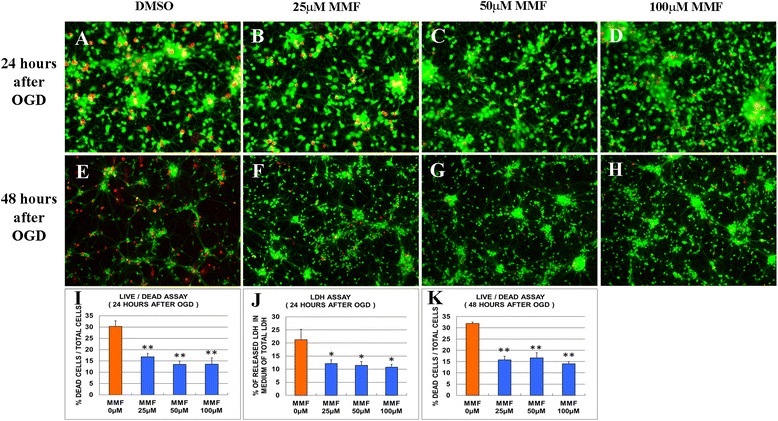
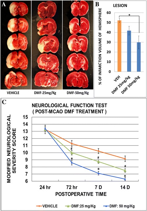
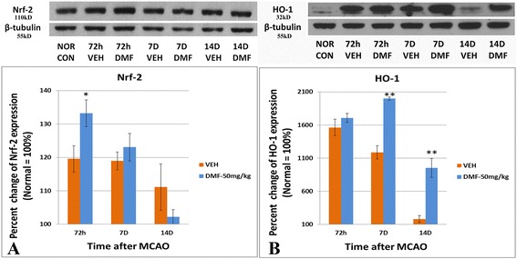
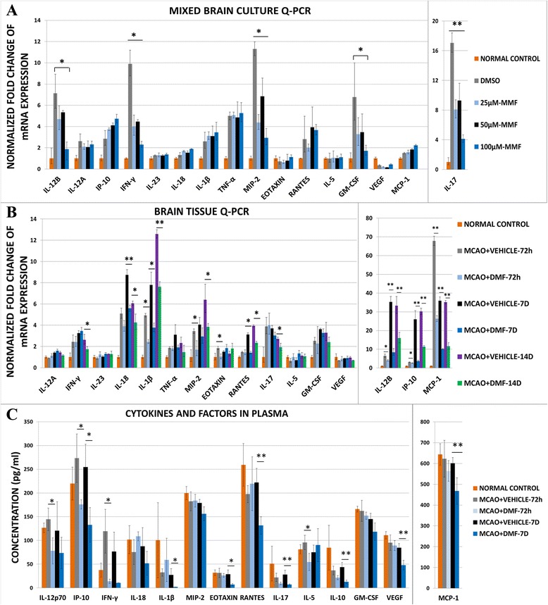
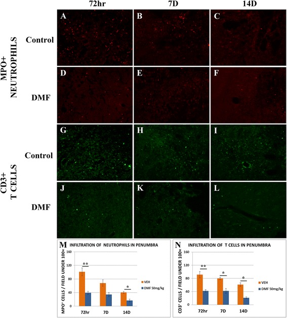
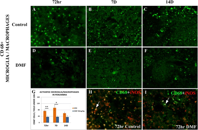
Results: We found that MMF significantly rescued cortical neurons from oxygen-glucose deprivation (OGD) in culture and suppressed pro-inflammatory cytokines produced by primary mixed neuron/glia cultures subjected to OGD. In rats, DMF treatment significantly decreased infarction volume by nearly 40 % and significantly improved neurobehavioral deficits after middle cerebral artery occlusion (MCAO). In the acute early phase (72 h after MCAO), DMF induced the expression of transcription factor Nrf2 and its downstream mediator HO-1, important for the protection of infarcted cells against oxidative stress. In addition to its antioxidant role, DMF also acted as a potent immunomodulator, reducing the infiltration of neutrophils and T cells and the number of activated microglia/macrophages in the infarct region by more than 50 % by 7-14 days after MCAO. Concomitantly, the levels of potentially harmful pro-inflammatory cytokines were greatly reduced in the plasma and brain and in OGD neuron/glia cultures.
Conclusions: We conclude that DMF is neuroprotective in experimental stroke because of its potent immunomodulatory and antioxidant effects and thus may be useful as a novel therapeutic agent to treat stroke in patients.
Keywords: Dimethyl fumarate; Inflammation; Stroke.
Figures
Similar articles
-
Protective Effect of GSK-3β/Nrf2 Mediated by Dimethyl Fumarate in Middle Cerebral Artery Embolization Reperfusion Rat Model.Curr Neurovasc Res. 2021;18(4):456-464. doi: 10.2174/1567202618666211109105024. Curr Neurovasc Res. 2021. PMID: 34751118
-
Dimethyl Fumarate and Monomethyl Fumarate Promote Post-Ischemic Recovery in Mice.Transl Stroke Res. 2016 Dec;7(6):535-547. doi: 10.1007/s12975-016-0496-0. Epub 2016 Sep 10. Transl Stroke Res. 2016. PMID: 27614618 Free PMC article.
-
Fumarate decreases edema volume and improves functional outcome after experimental stroke.Exp Neurol. 2017 Sep;295:144-154. doi: 10.1016/j.expneurol.2017.06.011. Epub 2017 Jun 8. Exp Neurol. 2017. PMID: 28602832
-
Mechanism of action and therapeutic potential of dimethyl fumarate in ischemic stroke.J Neurosci Res. 2023 Sep;101(9):1433-1446. doi: 10.1002/jnr.25202. Epub 2023 May 14. J Neurosci Res. 2023. PMID: 37183360 Review.
-
The inflammatory response in stroke.J Neuroimmunol. 2007 Mar;184(1-2):53-68. doi: 10.1016/j.jneuroim.2006.11.014. Epub 2006 Dec 26. J Neuroimmunol. 2007. PMID: 17188755 Free PMC article. Review.
Cited by
-
Emerging roles of GPR109A in regulation of neuroinflammation in neurological diseases and pain.Neural Regen Res. 2023 Apr;18(4):763-768. doi: 10.4103/1673-5374.354514. Neural Regen Res. 2023. PMID: 36204834 Free PMC article. Review.
-
3-n-butylphthalide exerts neuroprotective effects by enhancing anti-oxidation and attenuating mitochondrial dysfunction in an in vitro model of ischemic stroke.Drug Des Devel Ther. 2018 Dec 14;12:4261-4271. doi: 10.2147/DDDT.S189472. eCollection 2018. Drug Des Devel Ther. 2018. PMID: 30587922 Free PMC article.
-
Novel potential pharmacological applications of dimethyl fumarate-an overview and update.Front Pharmacol. 2023 Sep 7;14:1264842. doi: 10.3389/fphar.2023.1264842. eCollection 2023. Front Pharmacol. 2023. PMID: 37745068 Free PMC article. Review.
-
Neuroprotective effect of dimethyl fumarate in stroke: The role of nuclear factor erythroid 2-related factor 2.Iran J Neurol. 2019 Jul 6;18(3):108-113. Iran J Neurol. 2019. PMID: 31749931 Free PMC article.
-
Dimethyl Fumarate Limits Neuroinflammation and Oxidative Stress and Improves Cognitive Impairment After Polymicrobial Sepsis.Neurotox Res. 2018 Oct;34(3):418-430. doi: 10.1007/s12640-018-9900-8. Epub 2018 Apr 30. Neurotox Res. 2018. PMID: 29713994
References
-
- Global, regional, and national age–sex specific all-cause and cause-specific mortality for 240 causes of death, 1990–2013: a systematic analysis for the Global Burden of Disease Study 2013. Lancet [Internet]. 2014;385:117–71. Available from: http://www.pubmedcentral.nih.gov/articlerender.fcgi?artid=4340604&tool=p... [cited 2014 Dec 19] - PMC - PubMed
-
- Waltz AG. Pathophysiology of cerebral infarction. Clin Neurosurg [Internet] 1976;23:147–54. - PubMed
MeSH terms
Substances
LinkOut - more resources
Full Text Sources
Other Literature Sources
Medical
