. 2016 Nov;65(11):1657-1663.
doi: 10.1016/j.metabol.2016.08.008. Epub 2016 Sep 2.

Fat accumulation in the tongue is associated with male gender, abnormal upper airway patency and whole-body adiposity

Affiliations

Fat accumulation in the tongue is associated with male gender, abnormal upper airway patency and whole-body adiposity

Ivan R B Godoy et al. Metabolism. 2016 Nov.

Abstract

Objective: To examine associations between tongue adiposity with upper airway measures, whole-body adiposity and gender. We hypothesized that increased tongue adiposity is higher in males and positively associated with abnormal upper airway measures and whole-body adiposity.

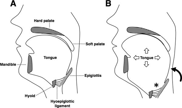
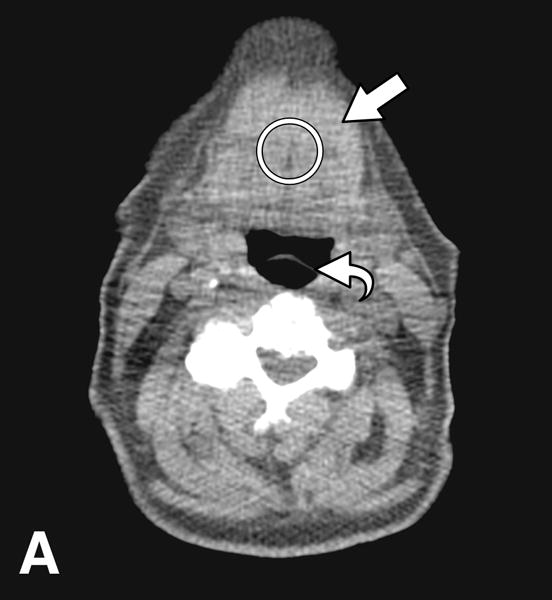
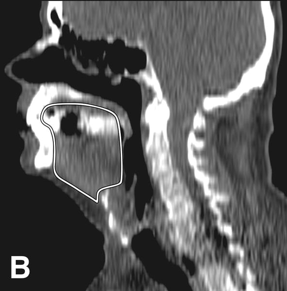
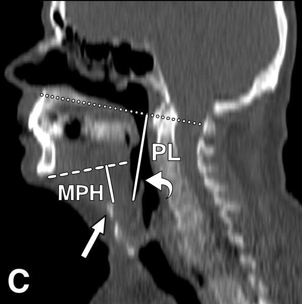
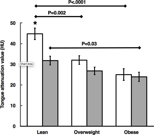
Methods: We studied subjects who underwent whole-body positron emission tomography/computed tomography to obtain tongue attenuation (TA) values and cross-sectional area, pharyngeal length (PL) and mandibular plane to hyoid distance (MPH), as well as abdominal circumference, abdominal subcutaneous and visceral (VAT) adipose tissue areas, neck circumference (NC) and neck adipose tissue area. Metabolic syndrome was determined from available clinical and laboratory data.

Results: We identified 206 patients (104 females, 102 males) with mean age 56±17years and mean body mass index (BMI) 28±6kg/m2 (range 16-47kg/m2). Males had lower TA values (P=0.0002) and higher upper airway measures (P<0.0001) independent of age and BMI (P<0.001). In all subjects, TA was negatively associated with upper airway measures (P<0.001). TA was negatively associated with body composition parameters (all P<0.0001), most notably with VAT (r=-0.53) and NC (r=-0.47). TA values were lower in subjects with metabolic syndrome (P<0.0001).

Conclusion: Increased tongue adiposity is influenced by gender and is associated with abnormal upper airway patency and body composition parameters.

Keywords: Body composition; Metabolic syndrome; Obesity; Tongue fat.


Conflict of interest statement

Disclosure: All authors have no conflicts of interest.

Figures

Figure 1
Figure 1
(A) The pharynx extends from the soft palate to the base of the epiglottis. The hyoid bone sits at the tongue base and is anchored by muscles and ligaments but does not articulate with other bones. (B) In obesity, tongue enlargement due to fat infiltration narrows the upper airway and displaces the hyoid caudally. The epiglottis follows the hyoid due to their connection via the hyoepiglottic ligament. Caudal displacement of the hyoid and epiglottis (*) leads to an increase in pharyngeal length (curved arrow). A longer pharynx is more collapsible, which predisposes to obstructive sleep apnea.
Figure 2
Figure 2
CT images from an obese male subject aged 74 years, with BMI 30 kg/m2, TA: 15.4 HU, tongue cross sectional area (CSA) 35.8 cm2, mandibular plane to hyoid distance (MPH) 2.9 cm, and pharyngeal length (PL): 7.2 cm. (A) Measurement of tongue attenuation values by placing a region of interest (circle) in the central portion of the tongue (arrow) on an axial slice obtained at the level of the epiglottis (curved arrow). (B) Sagittal CT image reconstruction through the midline showing tracing (white line) of tongue cross sectional area. (C) Sagittal CT image reconstruction through the midline demonstrating hard palate plane (dotted line) and epiglottis (curved arrow) providing PL, and the mandibular plane (dashed line) and hyoid bone (arrow) yielding MPH measures.
Figure 2
Figure 2
CT images from an obese male subject aged 74 years, with BMI 30 kg/m2, TA: 15.4 HU, tongue cross sectional area (CSA) 35.8 cm2, mandibular plane to hyoid distance (MPH) 2.9 cm, and pharyngeal length (PL): 7.2 cm. (A) Measurement of tongue attenuation values by placing a region of interest (circle) in the central portion of the tongue (arrow) on an axial slice obtained at the level of the epiglottis (curved arrow). (B) Sagittal CT image reconstruction through the midline showing tracing (white line) of tongue cross sectional area. (C) Sagittal CT image reconstruction through the midline demonstrating hard palate plane (dotted line) and epiglottis (curved arrow) providing PL, and the mandibular plane (dashed line) and hyoid bone (arrow) yielding MPH measures.
Figure 3
Figure 3
Mean differences in tongue adiposity per gender across BMI groups of lean, overweight, and obese subjects (females: white bars, males: gray bars). P values are for within-gender comparison across BMI categories using the Tukey-Kramer method. * denotes P=0.0004 for comparison between lean female versus male subjects by t test. Error bars denote ±standard error of mean.

References

    1. Young T, Peppard PE, Gottlieb DJ. Epidemiology of obstructive sleep apnea: a population health perspective. Am J Respir Crit Care Med. 2002;165:1217–39. - PubMed
    1. Heinzer R, Vat S, Marques-Vidal P, Marti-Soler H, Andries D, Tobback N, et al. Prevalence of sleep-disordered breathing in the general population: the HypnoLaus study. Lancet Respir Med. 2015;3:310–8. doi: 10.1016/S2213-2600(15)00043-0. - DOI - PMC - PubMed
    1. Schwab RJ, Gupta KB, Gefter WB, Metzger LJ, Hoffman EA, Pack AI. Upper airway and soft tissue anatomy in normal subjects and patients with sleep-disordered breathing. Significance of the lateral pharyngeal walls. Am J Respir Crit Care Med. 1995;152:1673–89. - PubMed
    1. Kim AM, Keenan BT, Jackson N, Chan EL, Staley B, Poptani H, et al. Tongue fat and its relationship to obstructive sleep apnea. Sleep. 2014;37:1639–48. doi: 10.5665/sleep.4072. - DOI - PMC - PubMed
    1. Brennick MJ, Delikatny J, Pack AI, Pickup S, Shinde S, Zhu JX, et al. Tongue fat infiltration in obese versus lean Zucker rats. Sleep. 2014;37:1095–102. 1102a–1102c. doi: 10.5665/sleep.3768. - DOI - PMC - PubMed

MeSH terms

LinkOut - more resources