Review
. 2017 Jan;101(1):77-86.
doi: 10.1189/jlb.2MR0416-201R. Epub 2016 Oct 12.

Cell-autonomous stress responses in innate immunity

Affiliations
Review

Cell-autonomous stress responses in innate immunity

Julien Moretti et al. J Leukoc Biol. 2017 Jan.

Abstract

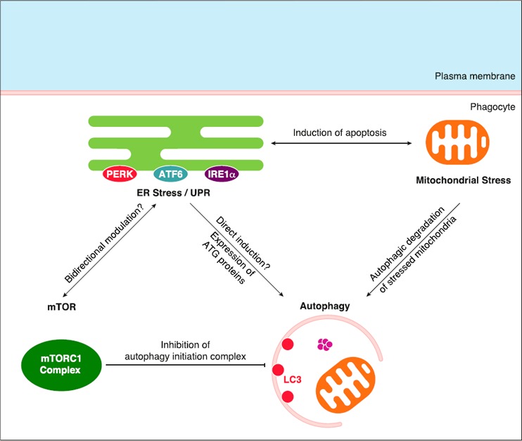
The innate immune response of phagocytes to microbes has long been known to depend on the core signaling cascades downstream of pattern recognition receptors (PRRs), which lead to expression and production of inflammatory cytokines that counteract infection and induce adaptive immunity. Cell-autonomous responses have recently emerged as important mechanisms of innate immunity. Either IFN-inducible or constitutive, these processes aim to guarantee cell homeostasis but have also been shown to modulate innate immune response to microbes and production of inflammatory cytokines. Among these constitutive cell-autonomous responses, autophagy is prominent and its role in innate immunity has been well characterized. Other stress responses, such as metabolic stress, the ER stress/unfolded protein response, mitochondrial stress, or the DNA damage response, seem to also be involved in innate immunity, although the precise mechanisms by which they regulate the innate immune response are not yet defined. Of importance, these distinct constitutive cell-autonomous responses appear to be interconnected and can also be modulated by microbes and PRRs, which add further complexity to the interplay between innate immune signaling and cell-autonomous responses in the mediation of an efficient innate immune response.

Keywords: DNA damage response; ER stress/UPR; autophagy; mTOR; mitochondrial stress; stress granules.


Figures

Figure 1
Figure 1
Autophagy is a defense mechanism against infection. After phagocytosis, invasive bacteria, such as S. typhimurium or Mycobacterium tuberculosis, escape the phagosome through the action of virulence factors. Once released into the cytoplasm, bacteria are targeted by the host ubiquitination machinery, which enables the interaction with autophagy receptors p62, NDP52, and optineurin and the recruitment of bacteria to preformed LC3‐labeled autophagosomes. Fusion of mature autophagosomes with lysosomes leads to autophagic degradation of bacteria.
Figure 2
Figure 2
Autophagy: induction by PRRs and inhibition of inflammasomes. PRRs, such as TLRs or NOD2, can induce autophagy via unknown molecular mechanisms. In response to diverse stimuli, NLRP3 and AIM2 inflammasomes are assembled and lead to production of proinflammatory cytokines IL‐1β and IL‐18. Autophagy is known to inhibit inflammasome activity. Inflammasome components NLRP3, AIM2, and ASC have been shown to be ubiquitinated (Ub) and degraded via autophagosomes, which explains inflammasome inhibition by autophagy.
Figure 3
Figure 3
The mTORC1 pathway: effect on TLR signaling cascades and modulation by bacteria. mTORC1 is activated primarily downstream of growth factor receptors (receptor tyrosine kinase) via the PI3K‐AKT‐TSC pathway or by amino acids in the cytoplasm. mTORC1 activates 4EBP1 and S6K, which promote protein translation. mTORC1 is also the main cytosolic inhibitor of autophagy. mTORC1 regulates TLR signaling via activation of MAPK and STAT3 or inhibition of NF‐κB. The mTORC1 pathway is modulated by pathogenic bacteria through the inhibition of AKT (in response to L. pneumophila) or amino acid starvation (in response to S. typhimurium). TRIF = TIR (Toll/IL‐1R) domain‐containing adapter inducing IFN‐β.
Figure 4
Figure 4
ER stress/UPR: effects on innate immunity. ER stress starts with the accumulation of unfolded proteins in the lumen of the ER. It activates a UPR via engagement of 3 UPR sensors and their respective signaling cascades: PERK‐EIF2α‐ATF4, ATF6, and IRE1α‐XBP‐1 pathways. These 3 UPR pathways lead to a global inhibition of protein translation and to expression of UPR target genes that are involved in the resolution of ER stress via protein folding processes or ER‐associated degradation of unfolded proteins (ERAD). ER stress/UPR was proposed to activate different immune signaling cascades, of note downstream of TLRs, such as JNK, NF‐κB, and IRF3 pathways, and, therefore, to promote the production of inflammatory cytokines. ER stress/UPR also activates NOD1/2‐dependent inflammatory cytokine production.
Figure 5
Figure 5
Connections between ER stress/UPR, mTORC1 pathway, autophagy, and mitochondrial stress. mTORC1 complex is known to inhibit autophagy via the inhibitory phosphorylation of components of the autophagy initiation complex. ER stress/UPR might promote autophagy directly or via expression of ATG proteins found among UPR target genes. Moreover, a bidirectional modulation seems to connect ER stress/UPR and the mTOR pathway, although no precise mechanism has been described. ER stress/UPR cooperates with mitochondrial stress (via Ca2+ flux and ROS production) to induce apoptosis. Stressed/damaged mitochondria are subjected to ubiquitination and recruitment to the autophagy machinery, which leads to their autophagic degradation.

References

    1. Janeway, C. A., Jr. (1989) Approaching the asymptote? Evolution and revolution in immunology. Cold Spring Harb. Symp. Quant. Biol. 54, 1–13. - PubMed
    1. Janeway, C. A., Jr. (1992) The immune system evolved to discriminate infectious nonself from noninfectious self. Immunol. Today 13, 11–16. - PubMed
    1. Thompson, M. R. , Kaminski, J. J. , Kurt‐Jones, E. A. , Fitzgerald, K. A. (2011) Pattern recognition receptors and the innate immune response to viral infection. Viruses 3, 920–940. - PMC - PubMed
    1. Moretti, J. , Blander, J. M. (2014) Insights into phagocytosis‐coupled activation of pattern recognition receptors and inflammasomes. Curr. Opin. Immunol. 26, 100–110. - PMC - PubMed
    1. MacMicking, J. D. (2012) Interferon‐inducible effector mechanisms in cell‐autonomous immunity. Nat. Rev. Immunol. 12, 367–382. - PMC - PubMed

Substances