. 2016 Oct 12;36(41):10529-10544.
doi: 10.1523/JNEUROSCI.1643-16.2016.

Neural Stem Cell Transplantation Induces Stroke Recovery by Upregulating Glutamate Transporter GLT-1 in Astrocytes

Affiliations

Neural Stem Cell Transplantation Induces Stroke Recovery by Upregulating Glutamate Transporter GLT-1 in Astrocytes

Marco Bacigaluppi et al. J Neurosci. .

Abstract

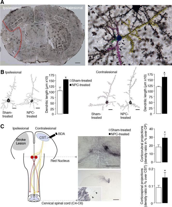
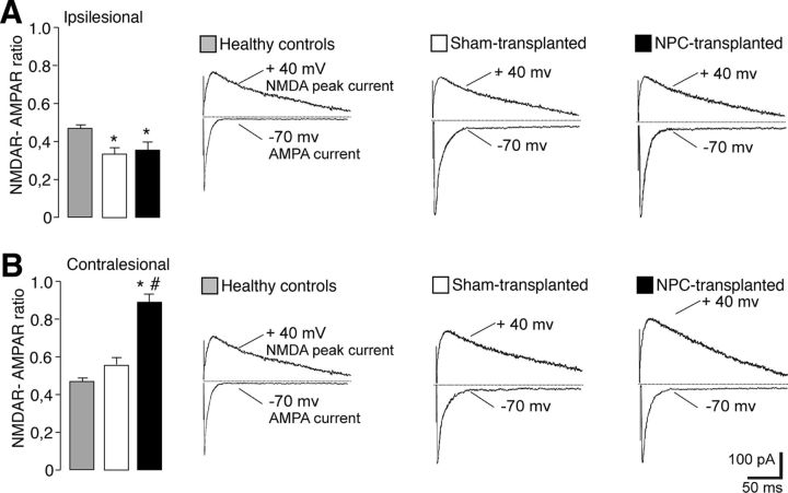
Ischemic stroke is the leading cause of disability, but effective therapies are currently widely lacking. Recovery from stroke is very much dependent on the possibility to develop treatments able to both halt the neurodegenerative process as well as to foster adaptive tissue plasticity. Here we show that ischemic mice treated with neural precursor cell (NPC) transplantation had on neurophysiological analysis, early after treatment, reduced presynaptic release of glutamate within the ipsilesional corticospinal tract (CST), and an enhanced NMDA-mediated excitatory transmission in the contralesional CST. Concurrently, NPC-treated mice displayed a reduced CST degeneration, increased axonal rewiring, and augmented dendritic arborization, resulting in long-term functional amelioration persisting up to 60 d after ischemia. The enhanced functional and structural plasticity relied on the capacity of transplanted NPCs to localize in the peri-ischemic and ischemic area, to promote the upregulation of the glial glutamate transporter 1 (GLT-1) on astrocytes and to reduce peri-ischemic extracellular glutamate. The upregulation of GLT-1 induced by transplanted NPCs was found to rely on the secretion of VEGF by NPCs. Blocking VEGF during the first week after stroke reduced GLT-1 upregulation as well as long-term behavioral recovery in NPC-treated mice. Our results show that NPC transplantation, by modulating the excitatory-inhibitory balance and stroke microenvironment, is a promising therapy to ameliorate disability, to promote tissue recovery and plasticity processes after stroke.

Significance statement: Tissue damage and loss of function occurring after stroke can be constrained by fostering plasticity processes of the brain. Over the past years, stem cell transplantation for repair of the CNS has received increasing interest, although underlying mechanism remain elusive. We here show that neural stem/precursor cell transplantation after ischemic stroke is able to foster axonal rewiring and dendritic plasticity and to induce long-term functional recovery. The observed therapeutic effect of neural precursor cells seems to underlie their capacity to upregulate the glial glutamate transporter on astrocytes through the vascular endothelial growth factor inducing favorable changes in the electrical and molecular stroke microenvironment. Cell-based approaches able to influence plasticity seem particularly suited to favor poststroke recovery.

Keywords: ischemia; neurophysiology; plasticity; recovery; stem cell; transplantation.


Figures

Figure 1.
Figure 1.
NPC treatment promotes postischemic neurological recovery and tissue protection. A, Experimental study design. B, Grip strength test of the right paretic forepaw and mNSS of sham-treated (white) and NPC-treated mice (black); n = 8 or 9 mice/group. C, Representative serial 3D reconstructions of the brains of a sham- and an NPC-treated mouse at 60 dpt. Yellow represents the ischemic lesion. Red represents the healthy striatum. Blue represents the lateral ventricles. Gray represents the contours of the hemispheres. Graphs on right represent the lesion volume (in mm3) and the atrophy of the ipsilesional hemisphere (as percentage of the contralateral hemisphere) (n = 4 or 5 mice/group). D, Quantification of the CST area evaluated on Bielschowsky-stained sections at level C4–C6 at 60 dpt (left) and quantification of the number of c-APP+ fibers in the ipsilesional CST at cervical spinal cord level (C4–C6) at 60 dpt in NPC-treated mice (right) (n = 4 mice/group). Representative microphotographs of a sham- and a NPC-treated mouse show c-APP+ fibers (green, arrowheads) of the ipsilesional CST, whereas the contralesional CST is denoted by the BDA (red; injected in the contralesional CST). Inset, Magnification showing c-APP+ fibers. Scale bar, 50 μm. Data are mean ± SEM. #p ≤ 0.05 (two-way ANOVA, Bonferroni post hoc test). ###p ≤ 0.001 (two-way ANOVA, Bonferroni post hoc test). *p ≤ 0.05, unpaired t test.
Figure 2.
Figure 2.
NPC-treated mice display increased dendritic plasticity and contralesional axonal sprouting. A, Representative coronal brain section of a Golgi-stained ischemic brain at 60 dpt. Red dashed line indicates the boundary of the ischemic lesion. White dashed boxes represent the fifth cortical layer region from which pyramidal neurons have been selected. Scale bar, 500 μm. Right, The dendritic tree (depicted in colors) of a pyramidal neuron traced by Neurolucida software. Scale bar, 10 μm. B, Representative reconstructions of Golgi-stained pyramidal neurons of the cortical layer V from sham- and NPC-treated mice at 60 dpt in the ipsilesional (left) and contralesional hemisphere (right). Scale bar, 10 μm. Graphs represent the integrated dendritic length of pyramidal neurons; n = 4 mice (8 neurons/animal per group). C, Schematic representation of the contralesional CST (gray and yellow lines, the latter with sprouting toward the lesioned side) examined by means of the anterograde tract-tracer BDA, injected into the contralesional motor cortex (left hemisphere). Axonal sproutings crossing the midline were examined at the level of the nucleus ruber (red circle; red represents rubrospinal tracts) and at the level of the cervical spinal cord enlargement C4–C6. Representative microphotographs illustrating BDA-traced corticorubral fibers (top) intersecting the midline (superimposed in white) between both red nuclei. Corticospinal fibers (bottom), at cervical spinal cord level (C4–C6), with fibers crossing the midline, are indicated by arrowheads in the magnified inset. Intersecting lines (white dashed lines) indicate the site where crossing fiber density was analyzed. Scale bars: top, 50 μm; bottom, 250 μm. Right, Graphs represent the quantitative analysis of BDA-labeled midline-crossing fibers at the level of red nucleus and cervical spinal cord (n = 4–8 mice/group). A–C, White bars represent sham-treated mice. Black bars represent NPC-treated mice. CP, Cortical peduncle. Data are mean ± SEM. *p ≤ 0.05 (t test).
Figure 3.
Figure 3.
NPC treatment rebalances the ipsilesional inhibitory and excitatory tone and promotes contralesional postsynaptic excitatory currents. Properties of excitatory (E) and inhibitory (I) spontaneous postsynaptic currents (s-PSC) recorded by patch clamp on acute tissue slices from ipsilesional peri-ischemic medium spiny striatal neurons (left panels) and contralesional MSNs (right panels) at 10 dpt. In the ipsilesional hemisphere: A, Spontaneous IPSC frequency and amplitude are decreased after ischemia. NPC transplantation induces an increase of sIPSCs in the ischemic striatum. B, Ipsilesional sEPSC frequency is increased upon ischemia and reduced by NPC treatment. The NMDA antagonist MK-801 reduces sEPSC frequency after MCAO but not in control group. NPC transplantation has no effect on overactive NMDA receptors. C, D, NPC transplantation has no effect on sEPSC kinetic properties. In the contralesional hemisphere: E, NPC transplantation has no effect on sIPSCs compared with sham-treated mice. Nevertheless, NPC transplantation reduces presynaptic sEPSCs (F) and potentiates NMDA-dependent postsynaptic sEPSCs (G, H). I, J, The electrophysiological traces are examples of sEPSCs (downward deflections) recorded from striatal neurons of a control, a sham-, and a NPC-treated animal in the ipsilesional (I) and contralesional (J) hemisphere. The effect of MK-801 on NPC-treated neurons is shown. Gray bars represent healthy controls. White bars represent sham-treated mice. Black bars represent NPC-treated mice. n = 3 or 4 mice/group, 4 or 5 neurons /hemisphere. Data are mean ± SEM. *p ≤ 0.05 versus healthy controls (one-way ANOVA, Tukey's post hoc test). #p ≤ 0.05 versus sham-treated mice (one-way ANOVA, Tukey's post hoc test).
Figure 4.
Figure 4.
NPC treatment enhances contralesional synaptic strength. Relative contribution of AMPARs and NMDARs to the eEPSCs expressed as ratio in the ipsilesional (A) and contralesional (B) striatum. The NMDAR/AMPAR current ratio for every patched neuron was computed in each condition. Representative electrophysiological traces show AMPAR-mediated and NMDAR-mediated eEPSCs, recorded from ipsilesional (A) and contralesional (B) striatal neurons, respectively, in the presence of 50 μm picrotoxin. NPC-transplanted (n = 7), sham-transplanted (n = 7), and healthy control mice (n = 11). Data are mean ± SEM. *p ≤ 0.05, compared with healthy controls (one-way ANOVA, Tukey HSD). #p ≤ 0.05, compared with sham-transplanted mice (one-way ANOVA, Tukey HSD).
Figure 5.
Figure 5.
Intravenously transplanted NPCs persist undifferentiated in the ischemic and peri-ischemic tissue. A, The distribution of transplanted NPCs localizing in the brain at 10 dpt is shown in the serial 3D brain reconstruction shown in a coronal view and in view from above. Green dots represent GFP+ NPCs. Yellow represents the ischemic lesion. Gray represents the contours of the hemispheres. B, Representative coronal brain section stained for GFP+ NPCs (in brown, highlighted by arrowheads in the magnified inset) showing at 10 dpt the transplanted NPCs localizing within the ischemic and peri-ischemic tissue. Nuclei counterstained by hematoxylin. C, Representative reconstruction displaying the localization of NPCs (GFP+ in green highlighted by white arrowheads) within the ischemic and peri-ischemic area (lesion border contoured by the dashed white line) in an NPC-treated mice at 10 dpt. Astrocytes are labeled by ALDH1L1 (red) and neurons by NeuN (white). Nuclei are counterstained with DAPI (blue). D, G, Representative microphotographs of transplanted NPCs localizing in the ischemic and peri-ischemic tissue. Most of the transplanted NPCs (GFP+, green in C–F, highlighted by white arrowheads) are found in close contact with inflammatory infiltrates composed of macrophages and microglia (F4/80+, red in D) and in close proximity to CD31+ vessels (blue in D, white in E). At 10 dpt, transplanted GFP+ NPCs mostly retain an undifferentiated phenotype and show only some neuroblast features (PSA-NCAM+, red in E). Even at 60 dpt, most of the GFP+ cells mostly exhibit undifferentiated features being negative for DCX (red in F), for ß-III-tubulin (white in F), and for GFAP+ (white in G). G, Dashed box is magnified in the marked contoured inset. Scale bars: A, 2 mm; B, 500 μm; C, 200 μm; D–G, 20 μm. Nuclei are counterstained with DAPI (white in D, blue in C, E–G). ALDH1L1, Aldehyde dehydrogenase 1 family member L1; β-III-tub, β-III-tubulin; PSA-NCAM, polysialylated neural cell adhesion molecule; CD31 [known also as platelet endothelial cell adhesion molecule (PECAM-1)]; DCX, doublecortin; F4/80 [also known as EGF-like module-containing mucin-like hormone receptor-like 1 (EMR1)]; NeuN, neuronal nuclei.
Figure 6.
Figure 6.
NPC treatment induces functional GLT-1 glutamate transporter upregulation in the peri-ischemic area. A, B, In vivo measurement of extracellular glutamate at 10 dpt by a microdialysis probe implanted in the striatum ipsilateral to the ischemic lesion. A, Representative cresyl violet-stained coronal ischemic brain section (0.2 mm from bregma) showing a sketched microdialysis probe implanted in the peri-ischemic striatum (ischemic lesion border in red). Note, in particular, the tip of the probe where black and white squares represent the active zone of the dialysis probe. The probe is perfused with either CSF at a rate of 2 μl/min (IN) and the perfusate is collected (OUT) every 10 min for glutamate measurement by high-performance liquid chromatography. Scale bar, 2 mm. B, Basal levels of glutamate in sham-treated and NPC-treated mice at 10 dpt. Shown is the average basal level of glutamate measured over 60 min (n = 6 or 7 mice per group). *p ≤ 0.05 (t test). C–E, Quantification of GLAST (C) and GLT-1 (D) expression in the peri-ischemic tissue over time revealed a significant increase of GLT-1 in NPC-treated animals at 10 dpt (n = 4–6 mice /group per time-point). Green line drawn on the schematic ischemic brain hemisphere visualizes the glial border where the quantification has been performed. ###p ≤ 0.001 (two-way ANOVA, Bonferroni post hoc test). E, Representative reconstructions of the peri-ischemic area (lesion border highlighted by the dashed white line) of a sham-treated and NPC-treated mouse showing the expression of GLT-1 (green) at 10 dpt. F, Representative immunofluorescence of an ischemic mouse at 10 dpt showing GLT-1 expression (red) specifically expressed on peri-ischemic astrocytes, stained with aquaporin 4 (AQP4, green). On the right of the merged image, single colored images (green and red channel) are shown. Nuclei are counterstained with DAPI (blue, E, F). G, Western blot for GLT-1 in ipsilesional striatum of sham-treated (representative image shown in left lane) and NPC-treated (representative image shown in right lane) mice at 10 dpt (n = 4 mice/group). *p ≤ 0.05 (t test). GLT-1 monomer (∼60 kDa) and dimer (∼120 kDa) were included in the densitometry analysis. Scale bars: E, 50 μm; F, 15 μm. Data are mean ± SEM. GLU, Glutamate.
Figure 7.
Figure 7.
GLT-1 inhibition in NPC-treated ischemic mice prevents functional amelioration. A, Timeline depicting the experimental design to block the overexpression of GLT-1 in NPC-treated mice. B, Grip strength test of the lesion-contralateral paretic forepaw and mNSS test showing over time progressive functional neurological improvement only in NPC-treated mice receiving intrastriatal (IS) PBS (NPC+PBSIS, black circle, n = 10 mice) compared with NPC-treated mice receiving DHK (NPC+DHKIS, black square, n = 11 mice) and sham-treated mice treated either with intrastriatal PBS (PBS+PBSIS, white up triangle, n = 5 mice) or DHK (PBS+DHKis, white down triangle, n = 10 mice). §p ≤ 0.05 (two-way ANOVA, Bonferroni post hoc test). C, Neurophysiological properties of sEPSCs recorded from contralesional striatal neurons after MCAO at 14 dpt. The effects of DHK injection in sham- and NPC-treated ischemic mice are shown. DHK injection blocked the NPC-induced increase of sEPSC duration and the NPC-induced sensitivity to NMDA antagonist MK801. *p ≤ 0.05 versus PBSIS-treated (one-way ANOVA, Tukey HSD). #p ≤ 0.05 versus healthy control (one-way ANOVA, Tukey HSD). n = 10–15 per group per condition. Data are mean ± SEM.
Figure 8.
Figure 8.
NPCs induce GLT-1 expression on astrocytes through the secretion of VEGF. Representation of the coculture setup: 5 × 10⁴ astrocytes (blue) are plated in the bottom well for 24 h either alone or in coculture with 3 × 10⁵ NPCs (green) seeded in the transwell. GLT-1 and GLAST expression evaluated on astrocytes alone or after coculture at (A) mRNA and (B) protein level. C, Quantification of Glt-1 and Glast mRNA expression on astrocytes alone or after coculture with NPCs after PI3 kinase inhibitor LY294002 (10 μm) treatment. D, The color-coded heatmaps represent the expression profile of selected growth factors on NPCs (top lanes) and of its receptors on astrocytes (bottom lanes) either alone or after coculture. Data are represented as ΔCt over GAPDH (note that a high ΔCt, displayed as colder colors, denotes a low expression profile). UN, Undetermined (white). Asterisks indicate the statistical significance by comparing monocultured to cocultured cells. E, ELISA for VEGF protein performed on cell medium derived from either NPCs alone or after coculture with astrocytes. F, Evaluation of Glt-1 and Glast expression at mRNA level on astrocytes after treatment with recombinant VEGF at increasing concentrations. G, Addition of the VEGF receptor inhibitor (676489, VEGFR2 kinase inhibitor IV, 500 nm, I-VEGFR) to astrocyte-NPC coculture inhibits Glt-1 upregulation on astrocytes. White bars represent GLT-1 expression. Black bars represent GLAST expression. B, E, Data are expressed as mean ± SEM. A, C, F, G, Data are in arbitrary units (AU). A, B, E, F, G, One of 2–5 independent experiments with n = 3–6 replicates per group. *p ≤ 0.05 (unpaired t test). **p ≤ 0.01 (unpaired t test). ***p ≤ 0.001 (unpaired t test). ****p ≤ 0.0001 (unpaired t test).
Figure 9.
Figure 9.
VEGF secretion by transplanted NPCs is necessary for GLT-1 upregulation and neurological recovery. A–C, Transplanted GFP+ NPCs (green, arrowhead) were found to be strongly positive for VEGF (red in A), but not for CNTF or BDNF (CNTF, red in B, BDNF, red in C). A, The cell denoted with the arrow is also shown in its x-z (lower edge) and y-z (right edge) projection, separated by the dashed line. B, C, βIII-tubulin axons are stained in white. Nuclei are counterstained by DAPI (blue). Scale bar, 25 μm. D, Quantification by ELISA of VEGF present in the ipsilesional and contralesional hemisphere of sham-treated and NPC-treated ischemic mice (n = 8 or 9 animals per group) at 10 dpt (sham or NPC treatment at 3 d after ischemia). E, Stereotactic injection of VEGF, but not of its diluent (PBS), in the striatum, enhances Glt-1 expression, while not affecting Glast. Glt-1 expression in mice injected with VEGF (2 μl into the left striatum, black bars) and with PBS (2 μl in the right striatum, white bars) 3 d after the injections (ratio of VEGF injected hemisphere over PBS injected hemisphere, n = 3 or 4 animals). F, Timeline depicting the experimental design to block the beneficial effect of NPC-secreted VEGF. A group of mice was subjected to histology at 10 dpt, whereas another group continued behavioral tests up to 60 d post-transplantation. G, Grip strength test and mNSS showing functional neurological improvement only in NPC-treated mice receiving the isotype antibody (NPC+ISO, black circle, n = 6 mice) compared with NPC-treated mice receiving VEGF neutralizing antibody (NPC+nAB-VEGF, black square, n = 7 mice), sham-treated mice either treated with isotype (PBS+ISO, white up triangle, n = 4 mice), or VEGF neutralizing antibody (PBS+nAB-VEGF, white down triangle, n = 8 mice). H, Quantification of GLT-1 protein expression in the peri-ischemic tissue at 10 dpt revealed that increase of GLT-1 induced by NPC-treated with isotype is significantly reduced in mice treated with NPC plus the VEGF neutralizing antibody (n = 4–6 mice/group). A, D, E, Data are mean ± SEM. B, Data are in arbitrary units (AU). *p ≤ 0.05 (t test). §p ≤ 0.05 (Dunn multiple comparison test). #p ≤ 0.05 (two-way ANOVA, Bonferroni post hoc test).

References

    1. Andres RH, Horie N, Slikker W, Keren-Gill H, Zhan K, Sun G, Manley NC, Pereira MP, Sheikh LA, McMillan EL, Schaar BT, Svendsen CN, Bliss TM, Steinberg GK. Human neural stem cells enhance structural plasticity and axonal transport in the ischaemic brain. Brain. 2011;134:1777–1789. doi: 10.1093/brain/awr094. - DOI - PMC - PubMed
    1. Bacigaluppi M, Pluchino S, Martino G, Kilic E, Hermann DM. Neural stem/precursor cells for the treatment of ischemic stroke. J Neurol Sci. 2008;265:73–77. doi: 10.1016/j.jns.2007.06.012. - DOI - PubMed
    1. Bacigaluppi M, Pluchino S, Peruzzotti-Jametti L, Kilic E, Kilic U, Salani G, Brambilla E, West MJ, Comi G, Martino G, Hermann DM. Delayed post-ischaemic neuroprotection following systemic neural stem cell transplantation involves multiple mechanisms. Brain. 2009;132:2239–2251. doi: 10.1093/brain/awp174. - DOI - PubMed
    1. Bavelier D, Levi DM, Li RW, Dan Y, Hensch TK. Removing brakes on adult brain plasticity: from molecular to behavioral interventions. J Neurosci. 2010;30:14964–14971. doi: 10.1523/JNEUROSCI.4812-10.2010. - DOI - PMC - PubMed
    1. Bernhardt J, Langhorne P, Lindley RI, Thrift AG, Ellery F, Collier J, Churilov L, Moodie M, Dewey H, Donnan G. Efficacy and safety of very early mobilisation within 24 h of stroke onset (AVERT): a randomised controlled trial. Lancet. 2015;386:46–55. doi: 10.1016/S0140-6736(15)60690-0. - DOI - PubMed

Publication types

MeSH terms

Substances