Review
. 2017 May;24(5):753-761.
doi: 10.1177/1933719116669056. Epub 2016 Oct 12.

Establishment of the Human Uteroplacental Circulation: A Historical Perspective

Affiliations
Review

Establishment of the Human Uteroplacental Circulation: A Historical Perspective

Kenna Degner et al. Reprod Sci. 2017 May.

Abstract

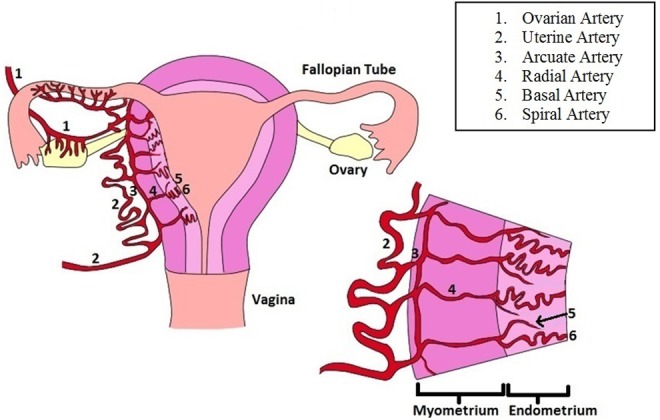
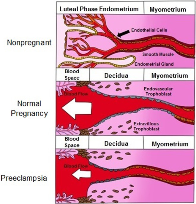
The uterine vasculature undergoes marked changes during pregnancy in order to provide the necessary increase in blood flow to support growth and nutrition of the uterus, placenta, and developing fetus. Pregnancy-associated uterine vascular transformations are orchestrated by a complex array of endocrine and cellular mechanisms to bring about structural modifications at the maternal-fetal interface, which collectively lead to development of the uteroplacental circulation. Understanding intrinsic uterine vascular remodeling in pregnancy is essential for understanding the physiologic and pathophysiologic regulation of maternal uterine blood flow. Aberrations of uterine vascular remodeling are potentially involved in the etiology of several pregnancy disorders, for example, preeclampsia, fetal growth restriction, and preterm labor; therefore, it is essential for subspecialist clinicians and investigators interested in reproductive physiology to fully understand the establishment of uteroplacental circulation. The foundational literature in this area is extensive; thus, a succinct review is likely to be a useful resource. Herein, we present and discuss a historical perspective on uterine vascular anatomy, maternal vascular growth associated with decidualization, trophoblast invasion, intervillous circulation, aberrations in uterine vascular modeling, and the clinical implications of improper development of the uteroplacental circulation.

Keywords: placenta; preeclampsia; pregnancy complications.


Conflict of interest statement

Declaration of Conflicting Interests: The author(s) declared no potential conflicts of interest with respect to the research, authorship, and/or publication of this article.

Figures

Figure 1.
Figure 1.
Uterine vascular anatomy: The uterine artery is the major vessel which provides blood to the uterus. At the surface of the uterus, the uterine artery branches into the arcuate artery. The radial artery, an offshoot of the arcuate artery, travels through the myometrium before giving off 2 branches at the myoendometrial junction: the basal artery and the spiral artery. Both the basal and spiral arteries transverse the endometrium and branch into capillary beds, but only the spiral artery reaches the luminal surface of the endometrium. The basal artery concludes at the basal surface of the endometrium.
Figure 2.
Figure 2.
Spiral artery remodeling in pregnancy: in the nonpregnant state, the spiral artery can be partitioned into 3 distinct layers: tunica intima, tunica media, and tunica adventitia. The vessel contains endothelial cells, shown in pink, and a smooth muscle layer, shown in red. In the pregnant state, the spiral artery retains only one of the original layers, the tunica adventitia. Endothelial cells are replaced by trophoblasts, shown in blue, and the layer of smooth muscle cells is also eliminated and supplanted by fibroblast cells and fibrinoid deposits which are shown in gray.
Figure 3.
Figure 3.
The intervillous circulation: Following pregnancy-associated vascular remodeling, the spiral arteries are known as the uteroplacental vessels. The uteroplacental vessels provide blood to the intervillous space, where the maternal blood is free to interact with the fetal villous tree. Although the term “cotyledon” has sometimes been used to refer to the incomplete partitions of the placenta, the human placenta is not truly cotyledonary and thus the term “functional cotyledon” is more appropriate.
Figure 4.
Figure 4.
Key events in the establishment of the uteroplacental circulation in normal and complicated pregnancies: In normal pregnancy (top), proper trophoblast invasion and vascular remodeling lay the foundation for a successful pregnancy. In pregnancies where these processes are interrupted or impaired (bottom) complications such as preeclampsia can occur.
Figure 5.
Figure 5.
Comparison of spiral artery remodeling in the nonpregnant, normal pregnant, and preeclamptic pregnant state: Normal, nonpregnant endometrium contains capillary beds and glands, which provide nourishment to the uterine endometrium. During pregnancy, these capillary beds and endometrial glands are destroyed, and the spiral arteries dilate and open directly into the intervillous space; thus, providing low-pressure, high-volume blood flow. In preeclampsia, some remodeling allows for a small degree of spiral artery dilation, but the volume of blood being delivered to the placenta is considerably less than in normal pregnancy.
Figure 6.
Figure 6.
Maternal spiral arteries which fail to undergo remodeling may demonstrate a vascular lesion known as acute atherosis. Lipid laden cells (blue arrow) and thickened, fibrinoid deposits in the vessel wall (black arrow) are characteristics of this lesion. Potential lipid containing macrophages (denoted by asterisk) are also apparent in this section.

References

    1. Intengan HD, Schiffrin EL. Vascular remodeling in hypertension: roles of apoptosis, inflammation, and fibrosis. Hypertension.2001;38(3 pt 2):581–587. - PubMed
    1. Mulvany MJ. Vascular remodelling of resistance vessels: can we define this? Cardiovasc Res. 1999;41(1):9–13. - PubMed
    1. Renna NF, de Las Heras N, Miatello RM. Pathophysiology of vascular remodeling in hypertension. Int J Hypertens. 2013;2013:808353. - PMC - PubMed
    1. Brosens I, Pijnenborg R, Vercruysse L, Romero R. The “Great Obstetrical Syndromes” are associated with disorders of deep placentation. Am J Obstet Gynecol. 2011;204(3):193–201. - PMC - PubMed
    1. Dixon HG, Robertson WB. A study of the vessels of the placental bed in normotensive and hypertensive women. J Obstet Gynaecol Br Emp. 1958;65(5):803–809. - PubMed

Publication types