. 2016 Oct 8:22:1156-1168.
eCollection 2016.

Proinflammatory cytokines decrease the expression of genes critical for RPE function

Affiliations

Proinflammatory cytokines decrease the expression of genes critical for RPE function

R Krishnan Kutty et al. Mol Vis. .

Abstract

Purpose: Proinflammatory cytokines interferon gamma (IFN-γ), tumor necrosis factor alpha (TNF-α), and interleukin-1 beta (IL-1β) secreted by infiltrating lymphocytes or macrophages may play a role in triggering RPE dysfunction associated with age-related macular degeneration (AMD). Binding of these proinflammatory cytokines to their specific receptors residing on the RPE cell surface can activate signaling pathways that, in turn, may dysregulate cellular gene expression. The purpose of the present study was to investigate whether IFN-γ, TNF-α, and IL-1β have an adverse effect on the expression of genes essential for RPE function, employing the RPE cell line ARPE-19 as a model system.

Methods: ARPE-19 cells were cultured for 3-4 months until they exhibited epithelial morphology and expressed mRNAs for visual cycle genes. The differentiated cells were treated with IFN-γ, TNF-α, and/or IL-1β, and gene expression was analyzed with real-time PCR analysis. Western immunoblotting was employed for the detection of proteins.

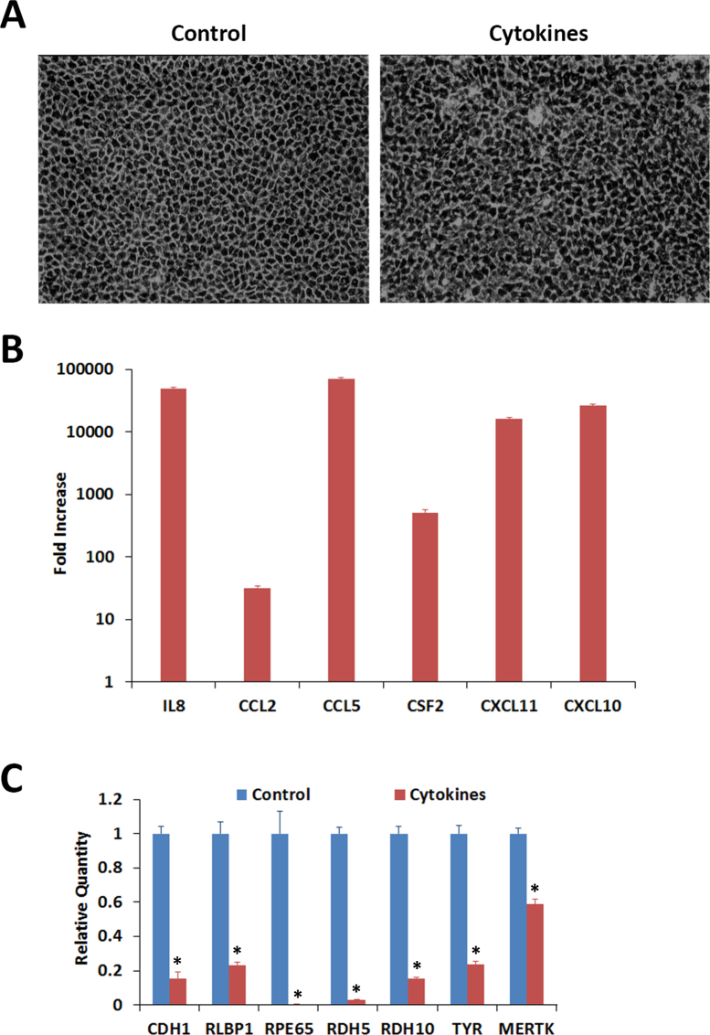
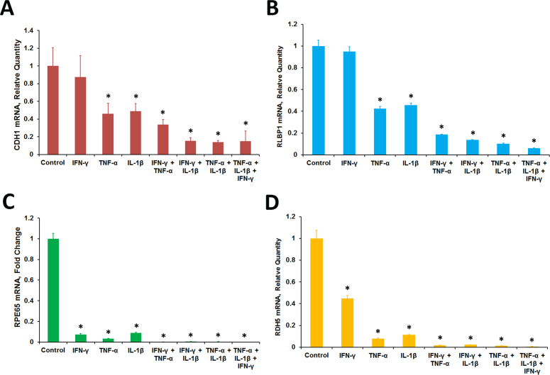
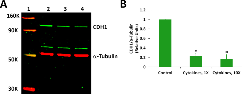
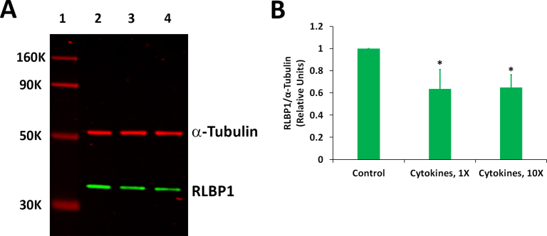
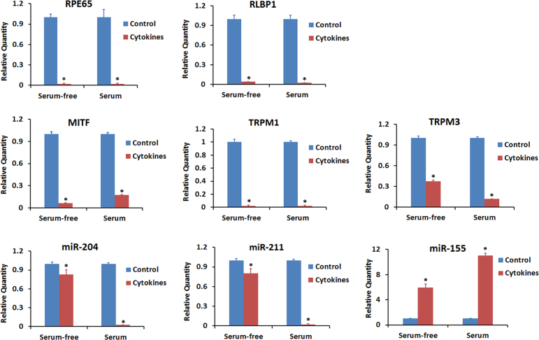
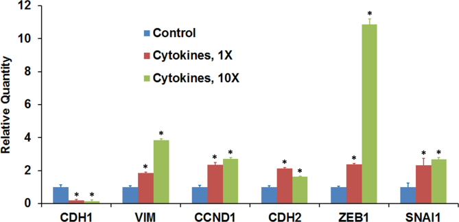
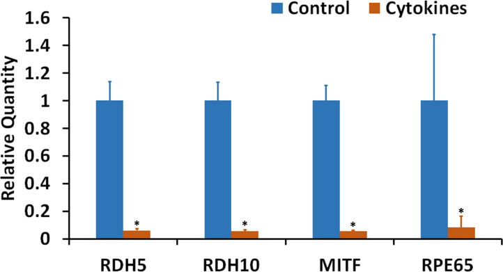
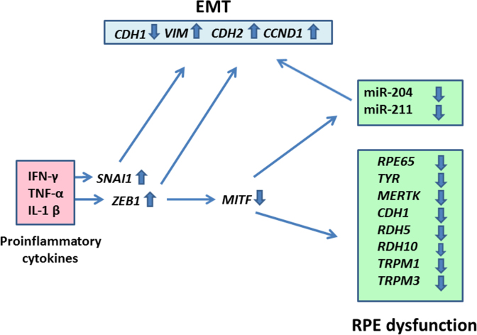
Results: Proinflammatory cytokines (IFN-γ + TNF-α + IL-1β) greatly increased the expression of chemokines and cytokines in cultured ARPE-19 cells that exhibited RPE characteristics. However, this response was accompanied by markedly decreased expression of genes important for RPE function, such as CDH1, RPE65, RDH5, RDH10, TYR, and MERTK. This was associated with decreased expression of the genes MITF, TRPM1, and TRPM3, as well as microRNAs miR-204 and miR-211, which are known to regulate RPE-specific gene expression. The decreased expression of the epithelial marker gene CDH1 was associated with increased expression of mesenchymal marker genes (CDH2, VIM, and CCND1) and epithelial-mesenchymal transition (EMT) promoting transcription factor genes (ZEB1 and SNAI1).

Conclusions: RPE cells exposed to proinflammatory cytokines IFN-γ, TNF-α, and IL-1β showed decreased expression of key genes involved in the visual cycle, epithelial morphology, and phagocytosis. This adverse effect of proinflammatory cytokines, which could be secreted by infiltrating lymphocytes or macrophages, on the expression of genes indispensable for RPE function may contribute to the RPE dysfunction implicated in AMD pathology.


Figures

Figure 1
Figure 1
Effect of proinflammatory cytokines on cultured ARPE-19 cells exhibiting RPE characteristics. The cells were treated with interferon gamma (IFN-γ; 10 u/ml), tumor necrosis factor alpha (TNF-α; 1 ng/ml), and interleukin-1 beta (IL-1β; 1 ng/ml) for 20 h in the absence of serum and their gene expression analyzed with real-time PCR. A: Control cells exhibited typical epithelial morphology when examined with phase contrast microscopy. Cells treated with cytokines appeared distorted with thickened cell junctions. Magnification: 100X. B: The cells responded to the proinflammatory cytokines by highly increasing the expression of cytokines and chemokines. Gene expression in the treated and control cells was analyzed with real-time PCR. The fold increases shown are statistically significant (p<0.05 compared to control, n = 4). C: The expression of the RPE characteristic genes when analyzed with real-time PCR decreased substantially in the differentiated ARPE-19 cells exposed to the proinflammatory cytokines. *p<0.5 when compared to control, n = 4.
Figure 2
Figure 2
Proinflammatory cytokines decreased the expression of RPE characteristic genes. Differentiated ARPE-19 cells were treated with interferon gamma (IFN-γ; 10 u/ml), tumor necrosis factor alpha (TNF-α; 1 ng/ml), interleukin-1 beta (IL-1β; ng/ml) either alone or in combination for 20 h in the absence of 1% serum. The gene transcripts were analyzed with real-time PCR. A: CDH1. B: RLBP1. C: RPE65. D: RDH5. *p<0.5 when compared to control, n = 4.
Figure 3
Figure 3
Proinflammatory cytokines decreased the expression of the CDH1 protein. Differentiated ARPE-19 cells were treated with the cytokines (1X = interferon gamma (IFN-γ), 10 u/ml + tumor necrosis factor alpha (TNF-α), 1 ng/ml + interleukin-1 beta (IL-1β), 1 ng/ml) for 4 days in the presence of 1% serum. A: Western immunoblot analysis of CDH1 expression. Lane 1: molecular weight markers; lane 2: control; lane 3: cytokines, 1X; and lane 4: cytokines, 10X. A representative blot from three similar experiments is shown. B: Histogram showing CDH1 expression. The CDH1 band intensity was normalized with corresponding α-tubulin band intensity; *p<0.05 when compared to control, n = 3.
Figure 4
Figure 4
Proinflammatory cytokines decreased the expression of the RLBP1 protein. Differentiated ARPE-19 cells were treated with the cytokines (1X = interferon gamma (IFN-γ), 10 u/ml + tumor necrosis factor alpha (TNF-α), 1 ng/ml + interleukin-1 beta (IL-1β), 1 ng/ml) for 4 days in the presence of 1% serum. A: Western immunoblot analysis of RLBP1 expression. Lane 1: molecular weight markers; lane 2: control; lane 3: cytokines, 1X; and lane 4: cytokines, 10X. A representative blot from three similar experiments is shown. B: Histogram showing RLBP1 expression. The RLBP1 band intensity was normalized with corresponding α-tubulin band intensity; *p<0.05 when compared to control, n = 3.
Figure 5
Figure 5
Proinflammatory cytokines decreased the expression of genes that control RPE function. Differentiated ARPE-19 cells were treated with the cytokines (interferon gamma (IFN-γ), 100 u/ml + tumor necrosis factor alpha (TNF-α), 10 ng/ml + interleukin-1 beta (IL-1β), 10 ng/ml) either for 20 h under serum-free conditions or for 4 days in the presence of 1% serum. Real-time PCR was employed to analyze the expression of the indicated transcripts and miRNAs. *p<0.05 when compared to respective control, n = 3.
Figure 6
Figure 6
Proinflammatory cytokines modulate the expression of genes that regulate the EMT. Differentiated ARPE-19 cells were treated with the cytokines (1X = interferon gamma (IFN-γ), 10 u/ml + tumor necrosis factor alpha (TNF-α), 1 ng/ml + interleukin-1 beta (IL-1β), 1 ng/ml) for 20 h in the absence of serum, and the expression transcripts for the epithelial marker gene (CDH1), mesenchymal marker genes (VIM, CCND1, and CDH2), and transcription factor genes (ZEB1 and SNAI1) was analyzed with real-time PCR. *p<0.5 when compared to control, n = 4.
Figure 7
Figure 7
Proinflammatory cytokines decreased the expression of RPE characteristic genes in an RPE cell culture system established from adult human donor eyes. The cultured cells were treated with the cytokines (interferon gamma (IFN-γ), 100 u/ml + tumor necrosis factor alpha (TNF-α), 10 ng/ml + interleukin-1 beta (IL-1β), 10 ng/ml) for 20 h under serum-free conditions. Real-time PCR analysis of the expression of mRNAs for RDH5, RDH10, MITF, and RPE65 is shown. *p<0.05 when compared to respective control, n = 4.
Figure 8
Figure 8
A schematic depiction of RPE cell dysfunction induced by IFN-γ, TNF-α, and IL-1β. The proinflammatory cytokines increased the expression of proepithelial–mesenchymal transition (EMT) transcription factor genes SNAI1 and ZEB1 in the RPE cells. This could result in the downregulation of the epithelial marker gene CDH1 and upregulation of the mesenchymal marker genes VIM, CDH2, and CCND1. Increased ZEB1 expression also caused a decrease in the expression of the MITF gene that encodes a transcription factor that controls RPE differentiation. This, in turn, may decrease the expression of the RPE functional genes RPE65, TYR, MERTK, RDH5, RDH10, TRPM1, and TRPM3. The decrease in MITF expression may also reduce the expression of miR-204 and miR-211, two microRNAs necessary for maintaining epithelial physiology and RPE function.

References

    1. Strauss O. The retinal pigment epithelium in visual function. Physiol Rev. 2005;85:845–81. - PubMed
    1. Sparrow JR, Hicks D, Hamel CP. The retinal pigment epithelium in health and disease. Curr Mol Med. 2010;10:802–23. - PMC - PubMed
    1. Buschini E, Piras A, Nuzzi R, Vercelli A. Age related macular degeneration and drusen: neuroinflammation in the retina. Prog Neurobiol. 2011;95:14–25. - PubMed
    1. Ambati J, Atkinson JP, Gelfand BD. Immunology of age-related macular degeneration. Nat Rev Immunol. 2013;13:438–51. - PMC - PubMed
    1. Cousins SW, Espinosa-Heidmann DG, Csaky KG. Monocyte activation in patients with age-related macular degeneration: a biomarker of risk for choroidal neovascularization? Arch Ophthalmol. 2004;122:1013–8. - PubMed

Publication types

MeSH terms