. 2016 Oct 6;1(16):e87157.
doi: 10.1172/jci.insight.87157.

IL-3 promotes the development of experimental autoimmune encephalitis

Affiliations

IL-3 promotes the development of experimental autoimmune encephalitis

Kerstin Renner et al. JCI Insight. .

Abstract

Little is known about the role of IL-3 in multiple sclerosis (MS) in humans and in experimental autoimmune encephalomyelitis (EAE). Using myelin oligodendrocyte glycoprotein (MOG) peptide-induced EAE, we show that CD4+ T cells are the main source of IL-3 and that cerebral IL-3 expression correlates with the influx of T cells into the brain. Blockade of IL-3 with monoclonal antibodies, analysis of IL-3 deficient mice, and adoptive transfer of leukocytes demonstrate that IL-3 plays an important role for development of clinical symptoms of EAE, for migration of leukocytes into the brain, and for cerebral expression of adhesion molecules and chemokines. In contrast, injection of recombinant IL-3 exacerbates EAE symptoms and cerebral inflammation. In patients with relapsing-remitting MS (RRMS), IL-3 expression by T cells is markedly upregulated during episodes of relapse. Our data indicate that IL-3 plays an important role in EAE and may represent a new target for treatment of MS.


Figures

Figure 1
Figure 1. MOG-specific expression of IL-3 by CD4+ T cells.
C57BL/6 (H-2b) mice were immunized with MOG peptide 35-55 on day 0. (A) Immediately before immunization (day 0) or 14 and 21 days after immunization (4–5 mice/time point), splenocytes were restimulated with MOG peptide 35-55 or PBS as control for 3 days, and the levels of IL-3, GM-CSF, and IFN-γ were measured in the supernatant. A pronounced MOG peptide 35-55–specific release of IL-3, GM-CSF, and IFN-γ was detectable 14 and 21 days after immunization. (B) Splenocytes obtained at day 14 after immunization were depleted of CD4+ or CD8+ T cells and restimulated with MOG peptide or PBS (3 mice/group). The MOG-specific release of IL-3 and GM-CSF was completely dependent on the presence of CD4+ T cells. (C) Before immunization (day 0) or 14 and 21 days after immunization, splenocytes were activated with PMA and ionomycin for 4 hours and stained for intracellular expression of IL-3, GM-CSF, IFN-γ, and IL-17 (5 mice/time point). The frequency of cytokine-positive CD4+ T cells markedly increased after immunization. One out of 2 representative experiments is shown. Data are represented as mean ±SEM, one-way ANOVA test of day 14 or day 21 vs. day 0: *P ≤ 0.05, **P ≤ 0.01, ***P < 0.001.
Figure 2
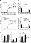
Figure 2. Blockade of IL-3 reduces development of EAE.
EAE was induced in C57BL/6 mice by immunization with MOG peptide 35-55 on day 0. From day 0–19, mice were treated with an intact or deglycosylated neutralizing anti–IL-3 antibody (anti–IL-3, 50 μg/day) or purified rat IgG (Control, 50 μg/day) (n = 14-15/group). (A and C) Clinical symptoms of EAE (EAE score) were significantly ameliorated (P < 0.01) in anti–IL-3–treated mice. (B and D) Leukocytes infiltrating the brain were quantified by flow cytometry on day 20. Monocytes (Monos) and total leukocytes (CD45+) were reduced by blockade of IL-3, while infiltrating CD4+ T cells, CD8+ T cells, and CD19+ B cells were not different between the groups. (E) On day 20, splenocytes were restimulated with MOG peptide 35-55 or PBS as control for 3 days, and the levels of IFN-γ, IL-6, and IL-17 were measured in the supernatant by ELISA. One out of 2 representative experiments is shown. Data are represented as mean ±SEM, Student’s t test of control vs. anti–IL-3: *P ≤ 0.05, **P < 0.01, ***P < 0.001.
Figure 3
Figure 3. Blockade of IL-3 prevents the early migration of leukocytes into the brain.
EAE was induced in C57BL/6 (H-2b) mice by immunization with MOG peptide 35-55 on day 0. From day 0–10, mice were treated with a neutralizing anti–IL-3 mAb (anti–IL-3, 50 μg/day) or purified rat IgG (Control, 50 μg/day) and analyzed on day 11. A third group of C57BL/6 (H-2b) mice was not immunized with MOG peptide 35-55 and not treated with antibodies (no EAE). (A) On day 11, leukocytes infiltrating the brain were quantified by flow cytometry (n = 9–10/group). Blockade of IL-3 reduced cerebral monocytes and total leukocytes (CD45+) by more than 50 %. Cerebral CD4+ and CD8+ T cells were reduced to the level of healthy nonimmunized mice. (B) Expression of E-selectin, P-selectin, RANTES (CCL5), and CXCL1 was quantified in the brain by qPCR in a separate experiment (n = 5-8/group). (C) Total splenocytes from C57BL/6 (H-2b) mice (800.000 cells/200 μl) were cultured for 24 or 48 hours with various cytokines (all 10 ng/ml). CCL5 was measured in the supernatant by ELISA. (D) Total splenocytes (Total) or splenocytes depleted of CD11b+ cells (11b) Ly6C+ cells (Ly6C) or CCR2+ cells (CCR2) (500.000 cells/200 μl) were cultured for 24 hours with IL-3 (10 ng/ml). CCL5 was measured in the supernatant by ELISA. One out of 2 representative experiments is shown. Data are represented as mean ±SEM, one-way ANOVA test of anti–IL-3 or no EAE vs. Control: *P ≤ 0.05, **P < 0.01, ***P < 0.001.
Figure 4
Figure 4. Adoptive transfer of CFSE-labeled leukocytes into mice with incipient EAE.
C57BL/6 (H-2b) mice were immunized with MOG peptide 35-55 on day 0 and treated from day 0–12 with a neutralizing anti–IL-3 antibody (anti–IL-3, 50 μg/day) or purified rat IgG (Control, 50 μg/day) (n = 7/group). On day 11, CFSE-labeled splenocytes were i.v. injected. These splenocytes were obtained on day 11 from C57BL/6 (H-2b) mice that were immunized with MOG peptide 35-55 on day 0 but not treated with mAbs. (A) Quantification of CFSE-labeled leukocytes in the brain and the spleen of recipients on day 13. Blockade of IL-3 significantly reduced the number of infiltrating CFSE+ T cells, B cells, and monocytes in the brain but not in the spleen. (B) Ratio of CFSE-labeled leukocytes detected within the brain and within the spleen. Monocytes migrated much more efficiently into the brain than T and B cells. Data are represented as mean ±SEM, Student’s t test of anti–IL-3 vs. Control: *P ≤ 0.05, **P < 0.01.
Figure 5
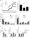
Figure 5. Reduced development of EAE in IL-3–deficient mice.
EAE was induced in C57BL/6 WT mice (IL-3+/+, n = 22), heterozygous IL-3–deficient mice (IL-3+/–, n = 18), and homozygous IL-3–deficient mice (IL-3–/–, n = 21) by immunization with MOG peptide 35-55 on day 0. (A) Clinical symptoms of EAE (EAE score) were significantly diminished in IL-3+/– and IL-3–/– mice, as seen by daily monitoring and by added scores (AUC). Pooled data of 2 independent experiments are shown. (B) Leukocytes were quantified in the brain and in the peripheral blood by flow cytometry on day 20 (n = 8–10/group). (C) On day 20, splenocytes were restimulated with MOG peptide 35-55 or PBS for 3 days, and the release of IL-3, GM-CSF, and IL-17 was measured in the supernatant by ELISA. Data are represented as mean ±SEM, one-way ANOVA test of IL-3+/– or IL-3–/– vs. IL-3+/+: *P ≤ 0.05, **P < 0.01, ***P < 0.001.
Figure 6
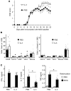
Figure 6. Injection of recombinant IL-3 exacerbates development of EAE.
EAE was induced in C57BL/6 (H-2b) mice by immunization with MOG peptide 35-55 on day 0. Mice were treated by daily i.p. injection of 200 ng recombinant murine IL-3 (IL-3) or PBS as control (PBS) from day 5–21 (n = 15/group). (A) Clinical symptoms of EAE (EAE score) are significantly higher in IL-3–treated mice. (B) Leukocyte subpopulations infiltrating the brain (left) and peripheral blood (right) were quantified by flow cytometry on day 22. (C) On day 22, splenocytes were restimulated with MOG peptide 35-55 or PBS as control for 3 days, and the level of IFN-γ, IL-17, and TNF were measured in the supernatant by ELISA. One out of 2 representative experiments is shown. Data are represented as mean ±SEM, Student’s t test of PBS vs. IL-3: *P ≤ 0.05.
Figure 7
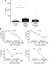
Figure 7. Expression of IL-3 in patients with MS and controls.
(A) PBMC of patients with RRMS with active disease (relapse) or nonactive disease (remission) and PBMC of controls were stimulated with anti-CD3. After 3 days, IL-3 was quantified in the supernatant by ELISA. RRMS patients with active MS (n = 19) showed increased IL-3 secretion compared with patients with nonactive MS (n = 30) and controls (n = 21). Data are represented as mean ±SEM. Student’s t test of controls vs. active MS. *P ≤ 0.05 (B) Four MS patients with active disease are depicted who stabilized over time and showed falling IL-3 values.

References

    1. Hofstetter HH, Karulin AY, Forsthuber TG, Ott PA, Tary-Lehmann M, Lehmann PV. The cytokine signature of MOG-specific CD4 cells in the EAE of C57BL/6 mice. J Neuroimmunol. 2005;170(1-2):105–114. doi: 10.1016/j.jneuroim.2005.09.004. - DOI - PubMed
    1. Karulin AY, Quast S, Hesse MD, Lehmann PV. Neuroantigen-Specific CD4 Cells Expressing Interferon-γ (IFN-γ), Interleukin (IL)-2 and IL-3 in a Mutually Exclusive Manner Prevail in Experimental Allergic Encephalomyelitis (EAE) Cells. 2012;1(3):576–596. - PMC - PubMed
    1. Tran EH, Prince EN, Owens T. IFN-gamma shapes immune invasion of the central nervous system via regulation of chemokines. J Immunol. 2000;164(5):2759–2768. doi: 10.4049/jimmunol.164.5.2759. - DOI - PubMed
    1. Lyons JA, Zhao ML, Fritz RB. Pathogenesis of acute passive murine encephalomyelitis II. Th1 phenotype of the inducing population is not sufficient to cause disease. J Neuroimmunol. 1999;93(1-2):26–36. doi: 10.1016/S0165-5728(98)00185-4. - DOI - PubMed
    1. Morris MM, Dyson H, Baker D, Harbige LS, Fazakerley JK, Amor S. Characterization of the cellular and cytokine response in the central nervous system following Semliki Forest virus infection. J Neuroimmunol. 1997;74(1-2):185–197. doi: 10.1016/S0165-5728(96)00786-2. - DOI - PubMed

Publication types