. 2016 Oct 6;1(16):e89362.
doi: 10.1172/jci.insight.89362.

Dual action of neurokinin-1 antagonists on Mas-related GPCRs

Affiliations

Dual action of neurokinin-1 antagonists on Mas-related GPCRs

Ehsan Azimi et al. JCI Insight. .

Abstract

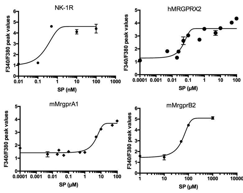
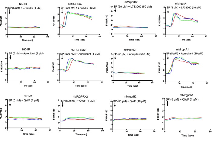
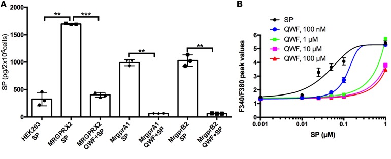
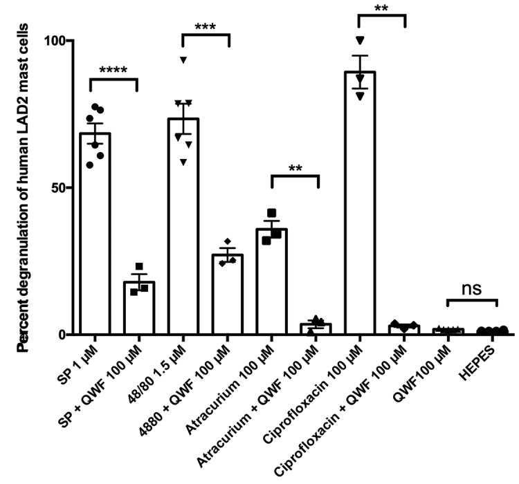
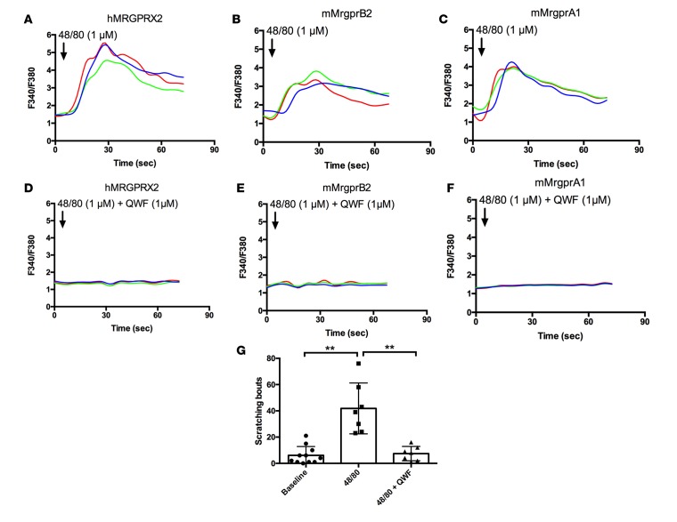
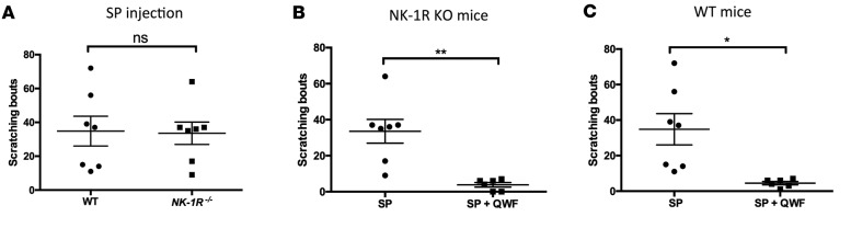
The challenge of translating findings from animal models to the clinic is well known. An example of this challenge is the striking effectiveness of neurokinin-1 receptor (NK-1R) antagonists in mouse models of inflammation coupled with their equally striking failure in clinical investigations in humans. Here, we provide an explanation for this dichotomy: Mas-related GPCRs (Mrgprs) mediate some aspects of inflammation that had been considered mediated by NK-1R. In support of this explanation, we show that conventional NK-1R antagonists have off-target activity on the mouse receptor MrgprB2 but not on the homologous human receptor MRGPRX2. An unrelated tripeptide NK-1R antagonist has dual activity on MRGPRX2. This tripeptide both suppresses itch in mice and inhibits degranulation from the LAD-2 human mast cell line elicited by basic secretagogue activation of MRGPRX2. Antagonists of Mrgprs may fill the void left by the failure of NK-1R antagonists.


Figures

Figure 1
Figure 1. Concentration-effect curves of substance P on NK-1R and Mrgprs.
HeLa cells were transfected with cDNAs encoding NK-1R, mouse MrgprB2, and mouse MrgprA1. A stably transfected HEK293 cell line was used for human MRGPRX2. Intracellular calcium ([Ca2+]i) was determined by ratiometric Fura-2 imaging after addition of substance P (SP). SP is known to activate NK-1R, human MRGPRX2, and mouse MrgprB2, as confirmed here. SP also activates mouse MrgprA1 but not several other mouse Mrgprs or human MRGPRX1, X3, and X4 (Supplemental Figure 1). The EC50s of SP for NK1-R, MRGPRX2, MrgprA1, and MrgprB2 are 0.5 nm, 100 nM, 5 μM, and 50 μM, respectively. These differential responses served to guide the concentrations of SP and antagonists used in the remainder of the studies. All data points are triplicates, and studies were performed at least twice.
Figure 2
Figure 2. NK-1R antagonists are selective antagonists of mouse and human Mrgprs.
L733060 (top row) and aprepitant (middle row) inhibit substance P–induced (SP-induced) activation of NK-1R and mouse MrgprB2 but do not inhibit human MRGPRX2 and mouse MrgprA1. QWF (bottom row) inhibits SP-induced activation of human MRGPRX2, mouse MrgprB2, and mouse MrgprA1. HeLa cells were transfected with cDNAs encoding NK-1R, mouse MrgprB2, and mouse MrgprA1. A stably transfected HEK293 cell line was used for human MRGPRX2. Intracellular calcium ([Ca2+]i) was determined by ratiometric Fura-2 imaging after addition of SP and antagonists. The concentration of SP for each receptor is based on the EC50s (Figure 1). The concentrations of the antagonists are used to balance out the concentration of SP. Each trace is a response from a different cell, and studies in each panel were performed at least twice.
Figure 3
Figure 3. The binding of substance P to Mrgprs is inhibited by QWF.
(A) ELISA was used to quantify the binding of substance P (SP) to nontransfected HEK293 cells and cells expressing human MRGPRX2, mouse MrgprB2, and mouse MrgprA1. SP binding is inhibited by QWF. P values for HEK293 SP vs. MRGPRX2 SP, MRGPRX2 SP vs. MRGPRX2 QWF+SP, HEK293 SP vs. MrgprB2 SP, MrgprB2 SP vs. MrgprB2 QWF+SP, HEK293 SP vs. MrgprA1 SP, and MrgprA1 SP vs. MrgprA1 QWF +SP are 0.0024, 0.0002, 0.0018, 0.0037, 0.0042, and 0.0012, respectively. HeLa cells were transfected with cDNAs encoding mouse MrgprB2 and mouse MrgprA1. A stably transfected HEK293 cell line was used for human MRGPRX2. *P ≤ 0.05, **P ≤ 0.01, ****P ≤ 0.001. (B) Concentration-effect curves with SP versus the indicated concentrations of QWF on a HEK293 cell line stably expressing human MRGPRX2. The results are consistent with QWF being a competitive antagonist of SP on human MRGPRX2. See Supplemental Figure 2 for the interaction of QWF with other GPCRs implicated in itch. The studies in A were performed 3 times and those in B were performed twice. 2-tailed unpaired Student’s t test was used.
Figure 4
Figure 4. The effect of QWF on IgE-independent mast cell degranulation.
The level of mast cell degranulation was assessed by the release of β-hexosaminidase in mast cell granules, as quantified by the level of its substrate, p-nitrophenyl N-acetyl-β-D-glucosamide (PNAG), digested in a colorimetric assay. QWF inhibits the degranulation induced by substance P (SP) (P < 0.0001), compound 48/80 (P = 0.0001), atracurium (P = 0.0025), and ciprofloxacin (P = 0.0039) in LAD2 mast cells. *P ≤ 0.05, **P ≤ 0.01, ***P ≤ 0.001, ****P ≤ 0.0001. These studies were performed 3 times using 2-tailed unpaired Student’s t test.
Figure 5
Figure 5. QWF is an antagonist of compound 48/80–induced Mrgpr activation and itch provocation.
(A–F) QWF inhibits activation of human MRGPRX2, mouse MrgprB2, and mouse MrgprA1 by compound 48/80. (G) Compound 48/80–induced itch is significantly decreased after coinjection with QWF (P = 0.002). Studies in AF were performed at least twice. The study in panel G was performed twice using 2-tailed unpaired Student’s t test was used. *P ≤ 0.05, **P ≤ 0.01.
Figure 6
Figure 6. Scratching from substance P is maintained in NK-1R–/– mice and is blocked by QWF.
(A) Itch induced by substance P (SP) (500 μM) is not decreased in NK-1R–/– mice compared with WT mice. (B) SP-induced itch is significantly decreased (P = 0.0040) in NK-1R–/– mice after coinjection with QWF (500 μM). (C) SP-induced itch is significantly decreased (P = 0.0134) in WT mice after coinjection with QWF (500 μM). These studies were performed twice using 2-tailed unpaired Student’s t test. *P ≤ 0.05, **P ≤ 0.01.

References

    1. Navari RM, Aapro M. Antiemetic prophylaxis for chemotherapy-induced nausea and vomiting. N Engl J Med. 2016;374(14):1356–1367. doi: 10.1056/NEJMra1515442. - DOI - PubMed
    1. Ramalho R, Soares R, Couto N, Moreira A. Tachykinin receptors antagonism for asthma: a systematic review. BMC Pulm Med. 2011;11:e89362. - PMC - PubMed
    1. May A, Goadsby PJ. Substance P receptor antagonists in the therapy of migraine. Expert Opin Investig Drugs. 2001;10(4):673–678. doi: 10.1517/13543784.10.4.673. - DOI - PubMed
    1. Rost K, Fleischer F, Nieber K. [Neurokinin 1 receptor antagonists--between hope and disappointment] Med Monatsschr Pharm. 2006;29(6):200–205. - PubMed
    1. Quartara L, Altamura M, Evangelista S, Maggi CA. Tachykinin receptor antagonists in clinical trials. Expert Opin Investig Drugs. 2009;18(12):1843–1864. doi: 10.1517/13543780903379530. - DOI - PubMed

Publication types

LinkOut - more resources