

Current Opinions and Areas of Consensus on the Role of the Cerebellum in Dystonia
- PMID: 27734238
- PMCID: PMC5336511
- DOI: 10.1007/s12311-016-0825-6
Current Opinions and Areas of Consensus on the Role of the Cerebellum in Dystonia
Abstract
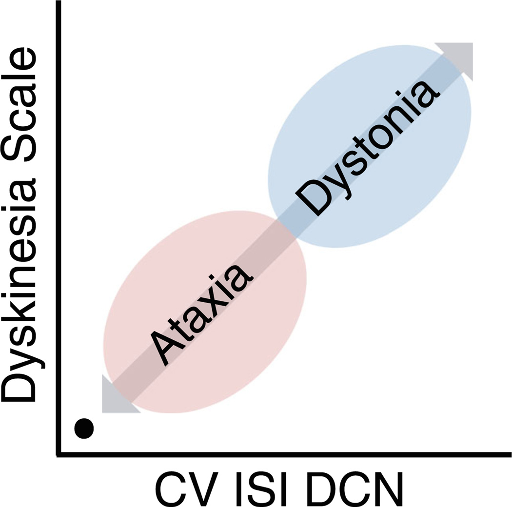
A role for the cerebellum in causing ataxia, a disorder characterized by uncoordinated movement, is widely accepted. Recent work has suggested that alterations in activity, connectivity, and structure of the cerebellum are also associated with dystonia, a neurological disorder characterized by abnormal and sustained muscle contractions often leading to abnormal maintained postures. In this manuscript, the authors discuss their views on how the cerebellum may play a role in dystonia. The following topics are discussed: The relationships between neuronal/network dysfunctions and motor abnormalities in rodent models of dystonia. Data about brain structure, cerebellar metabolism, cerebellar connections, and noninvasive cerebellar stimulation that support (or not) a role for the cerebellum in human dystonia. Connections between the cerebellum and motor cortical and sub-cortical structures that could support a role for the cerebellum in dystonia. Overall points of consensus include: Neuronal dysfunction originating in the cerebellum can drive dystonic movements in rodent model systems. Imaging and neurophysiological studies in humans suggest that the cerebellum plays a role in the pathophysiology of dystonia, but do not provide conclusive evidence that the cerebellum is the primary or sole neuroanatomical site of origin.
Keywords: Ataxia; Cerebellum; Circuits; DYT1; Dystonia; Networks.
Conflict of interest statement
Figures
References
-
- Bhatia KP, Marsden CD. The behavioral and motor consequences of focal lesions of the basal ganglia in man. Brain. 1994;117:859–876. - PubMed
-
- Malfait N, Sanger TD. Does dystonia always include co-contraction? A study of unconstrained reaching in children with primary and secondary dystonia. Exp Brain Res. 2007;176(2):206–216. - PubMed
Publication types
MeSH terms
Grants and funding
- P40 OD010996/OD/NIH HHS/United States
- R01 NS079750/NS/NINDS NIH HHS/United States
- P51 OD011132/OD/NIH HHS/United States
- R25 MH086466/MH/NIMH NIH HHS/United States
- R01 NS024328/NS/NINDS NIH HHS/United States
- R01 NS082296/NS/NINDS NIH HHS/United States
- P30 NS076405/NS/NINDS NIH HHS/United States
- R01 NS085054/NS/NINDS NIH HHS/United States
- R01 NS050808/NS/NINDS NIH HHS/United States
- U54 TR001456/TR/NCATS NIH HHS/United States
- R01 NS069936/NS/NINDS NIH HHS/United States
- U54 NS065701/NS/NINDS NIH HHS/United States
- R01 NS088528/NS/NINDS NIH HHS/United States
- K08 NS072158/NS/NINDS NIH HHS/United States
LinkOut - more resources
Full Text Sources
Other Literature Sources
Medical
