Review
. 2016 Oct;7(1):10.1128/ecosalplus.ESP-0006-2016.
doi: 10.1128/ecosalplus.ESP-0006-2016.

Animal Enterotoxigenic Escherichia coli

Affiliations
Review

Animal Enterotoxigenic Escherichia coli

J Daniel Dubreuil et al. EcoSal Plus. 2016 Oct.

Abstract

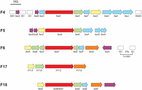
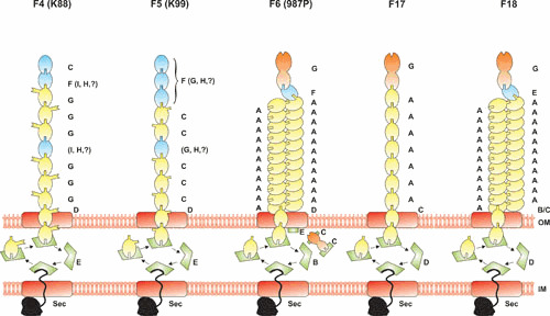
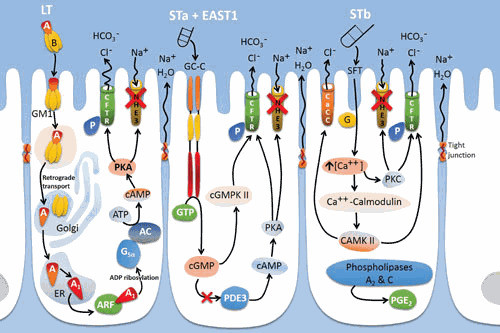
Enterotoxigenic Escherichia coli (ETEC) is the most common cause of E. coli diarrhea in farm animals. ETEC are characterized by the ability to produce two types of virulence factors: adhesins that promote binding to specific enterocyte receptors for intestinal colonization and enterotoxins responsible for fluid secretion. The best-characterized adhesins are expressed in the context of fimbriae, such as the F4 (also designated K88), F5 (K99), F6 (987P), F17, and F18 fimbriae. Once established in the animal small intestine, ETEC produce enterotoxin(s) that lead to diarrhea. The enterotoxins belong to two major classes: heat-labile toxins that consist of one active and five binding subunits (LT), and heat-stable toxins that are small polypeptides (STa, STb, and EAST1). This review describes the disease and pathogenesis of animal ETEC, the corresponding virulence genes and protein products of these bacteria, their regulation and targets in animal hosts, as well as mechanisms of action. Furthermore, vaccines, inhibitors, probiotics, and the identification of potential new targets by genomics are presented in the context of animal ETEC.


Figures

Figure 1
Figure 1
Genetic organization of animal ETEC fimbrial gene clusters. Genes encoding similar products or products with similar functions were labeled with the same pattern; genes for the major fimbrial subunit (yellow), minor fimbrial subunits (blue), minor adhesive subunit (orange), chaperones (green), usher (red), regulators (purple), and mobile or conjugation elements (white).
Figure 2
Figure 2
Fimbrial biogenesis models. The fimbriae all consist of the polymeric assembly of a major subunit (yellow) and of one or more minor subunits (blue), one of them being a tip adhesin (orange) for some fimbriae. For the K88 and K99 fimbriae, the major subunit is the adhesin. Usher proteins (red) locate in the outer membrane and channel the fimbrial subunits to the bacterial surface. All the fimbrial export systems use one periplasmic chaperone (green) for all the subunits, with the exception of the F6 fimbriae that have three chaperones, two being dedicated to two different fimbrial subunits. All the fimbrial proteins cross the inner membrane by using the general secretion (Sec) pathway (black), with the exception of fimbriae-specific regulators that remain in the cytoplasm (not shown).
Figure 3
Figure 3
Mechanism of action of ETEC toxins on intestinal epithelial cells. Signaling leading to water and electrolyte loss through activation of ion channels and loosening of tight junctions by the various toxins is described. CFTR, cystic fibrosis transmembrane regulator; AC, adenylate cyclase; ARF, ADP-ribosylation factor; PKA, protein kinase C; PKC, protein kinase C; GM1, ganglioside GM1; GC-C, guanylate cycles C; SFT, sulfatide; ER, endoplasmic reticulum; Gsα, α component of an heterotrimeric G protein; NHE3, Na+/H+-exchanger 3; PDE3, phosphodiesterase 3; cGMPKII, cGMP-dependent protein kinase II; cAMPKII, calmodulin-dependent protein kinase II; CaCC, calcium-activated chloride channel; P, phosphorylation.

References

    1. Nagy B, Fekete PZ. 2005. Enterotoxigenic Escherichia coli in veterinary medicine. Int J Med Microbiol 295:443–454. [PubMed]10.1016/j.ijmm.2005.07.003 - DOI - PubMed
    1. Kopic S, Geibel JP. 2010. Toxin mediated diarrhea in the 21 century: the pathophysiology of intestinal ion transport in the course of ETEC, V. cholerae and rotavirus infection. Toxins (Basel) 2:2132–2157. [PubMed]10.3390/toxins2082132 - DOI - PMC - PubMed
    1. Dubreuil JD. 2012. The whole Shebang: the gastrointestinal tract, Escherichia coli enterotoxins and secretion. Curr Issues Mol Biol 14:71–82. [PubMed] - PubMed
    1. van Beers-Schreurs HM, Vellenga L, Wensing T, Breukink HJ. 1992. The pathogenesis of the post-weaning syndrome in weaned piglets: a review. Vet Q 14:29–34. [PubMed]10.1080/01652176.1992.9694322 - DOI - PubMed
    1. Fairbrother JM, Nadeau E, Gyles CL. 2005. Escherichia coli in postweaning diarrhea in pigs: an update on bacterial types, pathogenesis, and prevention strategies. Anim Health Res Rev 6:17–39. [PubMed]10.1079/AHR2005105 - DOI - PubMed

Publication types

MeSH terms