

An Infrared Absorbance Sensor for the Detection of Melanoma in Skin Biopsies
- PMID: 27735858
- PMCID: PMC5087447
- DOI: 10.3390/s16101659
An Infrared Absorbance Sensor for the Detection of Melanoma in Skin Biopsies
Abstract
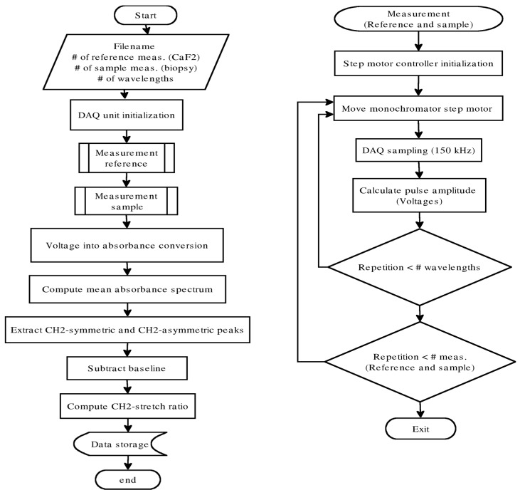
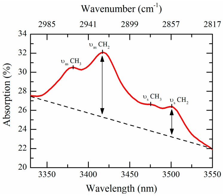
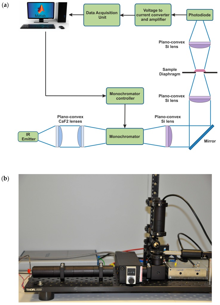
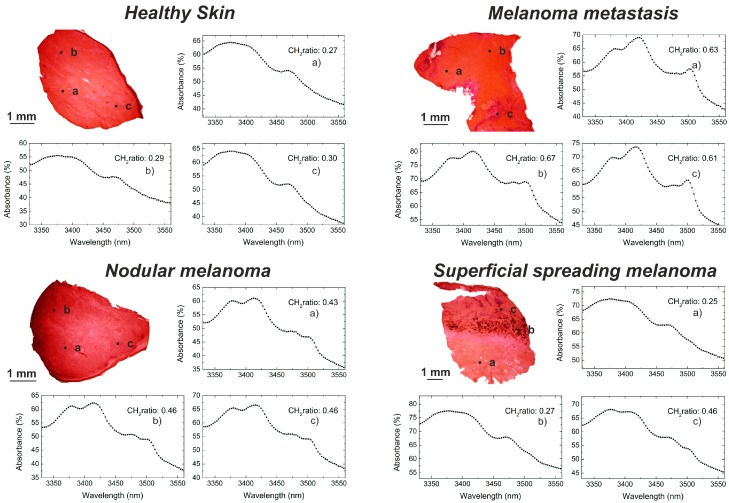
An infrared (IR) absorbance sensor has been designed, realized and tested with the aim of detecting malignant melanomas in human skin biopsies. The sensor has been designed to obtain fast measurements (80 s) of a biopsy using a small light spot (0.5 mm in diameter, typically five to 10 times smaller than the biopsy size) to investigate different biopsy areas. The sensor has been equipped with a monochromator to record the whole IR spectrum in the 3330-3570 nm wavelength range (where methylene and methyl stretching vibrations occur) for a qualitative spectral investigation. From the collected spectra, the CH₂ stretch ratio values (ratio of the absorption intensities of the symmetric to asymmetric CH₂ stretching peaks) are determined and studied as a cancer indicator. Melanoma areas exhibit different spectral shapes and significantly higher CH₂ stretch ratios when compared to healthy skin. The results of the infrared investigation are compared with standard histology. This study shows that the IR sensor is a promising supportive tool to improve the diagnosis of melanoma during histopathological analysis, decreasing the risk of misdiagnosis.
Keywords: absorbance spectroscopy; infrared sensor; melanoma; skin biopsy.
Conflict of interest statement
The authors declare no conflict of interest.
Figures
Similar articles
-
FT-IR Spectroscopy Study in Early Diagnosis of Skin Cancer.In Vivo. 2017 Nov-Dec;31(6):1131-1137. doi: 10.21873/invivo.11179. In Vivo. 2017. PMID: 29102935 Free PMC article.
-
Discriminating nevus and melanoma on paraffin-embedded skin biopsies using FTIR microspectroscopy.Biochim Biophys Acta. 2005 Aug 5;1724(3):262-9. doi: 10.1016/j.bbagen.2005.04.020. Biochim Biophys Acta. 2005. PMID: 15935560
-
Infrared and Raman Spectroscopic Studies of Molecular Disorders in Skin Cancer.In Vivo. 2019 Mar-Apr;33(2):567-572. doi: 10.21873/invivo.11512. In Vivo. 2019. PMID: 30804143 Free PMC article.
-
Role of In Vivo Reflectance Confocal Microscopy in the Analysis of Melanocytic Lesions.Acta Dermatovenerol Croat. 2018 Apr;26(1):64-67. Acta Dermatovenerol Croat. 2018. PMID: 29782304 Review.
-
Malignant Melanoma: Beyond the Basics.Plast Reconstr Surg. 2016 Aug;138(2):330e-340e. doi: 10.1097/PRS.0000000000002367. Plast Reconstr Surg. 2016. PMID: 27465194 Review.
Cited by
-
Skin cancer detection using non-invasive techniques.RSC Adv. 2018 Aug 6;8(49):28095-28130. doi: 10.1039/c8ra04164d. eCollection 2018 Aug 2. RSC Adv. 2018. PMID: 35542700 Free PMC article. Review.
-
Optical Technologies for the Improvement of Skin Cancer Diagnosis: A Review.Sensors (Basel). 2021 Jan 2;21(1):252. doi: 10.3390/s21010252. Sensors (Basel). 2021. PMID: 33401739 Free PMC article. Review.
-
Performance Assessment of Low-Cost Thermal Cameras for Medical Applications.Sensors (Basel). 2020 Feb 28;20(5):1321. doi: 10.3390/s20051321. Sensors (Basel). 2020. PMID: 32121299 Free PMC article.
-
Electromagnetic Sensing Techniques for Monitoring Atopic Dermatitis-Current Practices and Possible Advancements: A Review.Sensors (Basel). 2023 Apr 12;23(8):3935. doi: 10.3390/s23083935. Sensors (Basel). 2023. PMID: 37112275 Free PMC article. Review.
References
MeSH terms
LinkOut - more resources
Full Text Sources
Other Literature Sources
Medical
