. 2016 Oct 12;20(4):493-503.
doi: 10.1016/j.chom.2016.09.004.

Enterococcal Metabolite Cues Facilitate Interspecies Niche Modulation and Polymicrobial Infection

Affiliations

Enterococcal Metabolite Cues Facilitate Interspecies Niche Modulation and Polymicrobial Infection

Damien Keogh et al. Cell Host Microbe. .

Abstract

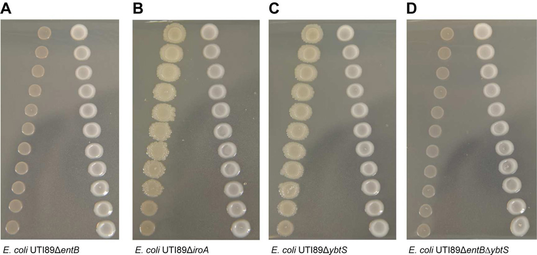
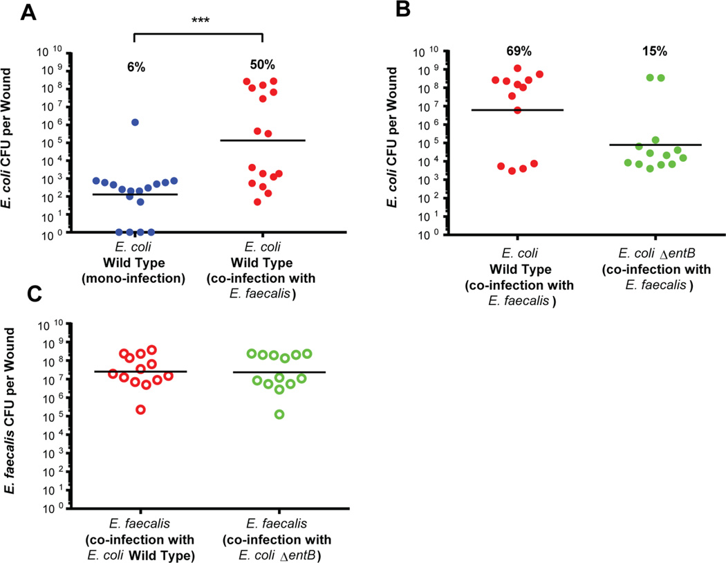
Enterococcus faecalis is frequently associated with polymicrobial infections of the urinary tract, indwelling catheters, and surgical wound sites. E. faecalis co-exists with Escherichia coli and other pathogens in wound infections, but mechanisms that govern polymicrobial colonization and pathogenesis are poorly defined. During infection, bacteria must overcome multiple host defenses, including nutrient iron limitation, to persist and cause disease. In this study, we investigated the contribution of E. faecalis to mixed-species infection when iron availability is restricted. We show that E. faecalis significantly augments E. coli biofilm growth and survival in vitro and in vivo by exporting L-ornithine. This metabolic cue facilitates E. coli biosynthesis of the enterobactin siderophore, allowing E. coli growth and biofilm formation in iron-limiting conditions that would otherwise restrict its growth. Thus, E. faecalis modulates its local environment by contributing growth-promoting cues that allow co-infecting organisms to overcome iron limitation and promotes polymicrobial infections.

Keywords: Enterococcus faecalis; Escherichia coli; iron; nutritional immunity; polymicrobial infection; wound infection.


Figures

Figure 1
Figure 1. E. faecalis promotes E. coli biofilm growth under iron limitation
(A–B) E. coli, E. faecalis, Mix(1Ec:1Ef), Mix(1Ec:4Ef) and Mix(1Ec:19Ef) biofilm growth in 22D supplemented TSBG. (C) Supplementation with 50µM FeCl3 restored biomass to iron replete levels. (A–C) Representative macrocolony images at 120hr; scale bars represent 1cm. (D & E) Time course enumeration of E. coli and E. faecalis, respectively, from macrocolonies with single or mixed inoculums under iron limitation. (F) E. coli, E. faecalis and Mix(1Ec:19Ef) biofilm biomass in TSBG supplemented with 22D. Statistical significance was determined by Two-Way ANOVA with Tukey’s test for multiple comparisons, **** P<0.0001. (G) Enumeration of planktonic growth at 120hr for E. coli, E. faecalis and Mix(1Ec:19Ef) in TSBG with 0.1, 0.2 and 0.7mM 22D. (D–G) n = 3 with three technical replicates.
Figure 2
Figure 2. E. faecalis proximity causes growth and transcriptome changes in E. coli
(A) Proximity assay of E. coli and E. faecalis (120hr) macrocolonies respectively in TSBG supplemented with 22D, inoculated from 1cm (proximal) to 5cm (distal). Scale bar represents 1cm. Black brackets indicate proximal and distal sample sites. (B) Enumeration at 24hr and 120hr for E. coli and E. faecalis at both proximal and distal sites, n = 3 with three technical replicates. (C) Statistical enrichment analysis of statistically significant E. coli pathways within top 100 & 500 differentially regulated genes (Benjamini-Hochberg adjusted P<0.05). KEGGid: pathway identifier used by KEGG; adjP: Benjamini- Hochberg corrected P-value; Ngenes: number of upregulated genes that are member of the pathway; EF: enrichment factor, the quotient of the observed proportion of pathway annotated genes to the expected proportion under the null hypothesis of no association. (D,E,F) Transcriptional gene expression profiling of E. coli in proximity to E. faecalis. Differentially regulated genes and functional categories (≥2-fold change, FDR<0.05) for (C) E. coli, (E) for E. coli iron related systems, and (F) for E. faecalis when grown in close proximity to E. coli. The black bar indicates the median value for each functional category; each circle represents one gene with red color indicating a functional category where the median value represents decreased expression, and green indicating increased expression; n = 3 biological replicates.
Figure 3
Figure 3. E. coli enterobactin siderophore mutants do not undergo E. faecalis-mediated growth enhancement
Proximity assays of 120hr macrocolonies in TSBG supplemented with 22D with E. faecalis and (A) E. coli UTI89ΔentB, (B) E. coli UTI89ΔiroA, (C) E. coli UTI89ΔybtS, and (D) E. coli UTI89ΔentBΔybtS.
Figure 4
Figure 4. E. faecalis co-infection increases the growth of E. coli in a mouse model of wound infection
Mouse wound infection with bacterial burdens determined at 24hpi. The E. coli inoculum in single species controls and/or mixed species infections was 2–4×10²CFU/wound for E. coli and 2–4×10⁶CFU/wound. (A) 24hpi E. coli CFU for mono-infected and co-infected wounds. (B) 24hpi E. coli wild type CFU for co-infected compared to E. coli UTI89ΔentB co-infected wounds. (C) E. faecalis CFU at 24hpi from wounds co-infected with E. coli wild type or E. coli UTI89ΔentB. Recovered titers of zero were set to the limit of detection of the assay for statistical analyses and graphical representation in all figures. Horizontal bars represent the median value for each group of mice. N = 3 biological replicates. Statistical significance was determined by the Mann-Whitney test with Dunn’s post-test for multiple comparisons, *** P=0.0009. Percentages indicate the proportion of data points falling above 1×10⁵CFU/wound for each group.
Figure 5
Figure 5. L-ornithine Supplementation Enhances E. coli wild type biofilm but not E. coli UTI89ΔentB under iron limitation
(A–B) E. coli wild type and ΔentB biofilm respectively at 120hr in TSBG supplemented with 22D. L-ornithine supplementation is indicated by the black bar with concentration indicated above. Representative data from three independent experiments is shown, where the trend is consistent among all. Statistical significance of the L-ornithine supplemented samples compared to the non-supplemented E. coli control was determined by Two-Way ANOVA with Tukey’s test for multiple comparisons, * P≤0.05, ** P≤0.01, *** P≤0.001, **** P≤0.0001.
Figure 6
Figure 6. Model for E. faecalis Modulation of the Polymicrobial Infection Niche
(A) Schematic representation of the arginine biosynthesis pathway. Genes encoding the enzymes involved at each step are italicized. Metabolites detected via non-targeted metabolomics are highlighted in green. (B) Model of a polymicrobial infection niche where E. faecalis (blue) modulates E. coli (green) through amino acid flux (ornithine). Siderophore biosynthesis and acquisition are indicated by green arrows. Host iron-bound proteins (from serum and intracellular sources) are highlighted red. The model depicts transition from the initial colonization stage with low E. coli titers to proliferation and infection at higher E. coli titers.

Comment in

References

    1. Archibald F. Manganese: its acquisition by and function in the lactic acid bacteria. Crit Rev Microbiol. 1986;13:63–109. - PubMed
    1. Barcelona-Andres B, Marina A, Rubio V. Gene Structure, Organization, Expression, and Potential Regulatory Mechanisms of Arginine Catabolism in Enterococcus faecalis. Journal of Bacteriology. 2002;184:6289–6300. - PMC - PubMed
    1. Bourgogne A, Hilsenbeck SG, Dunny GM, Murray BE. Comparison of OG1RF and an isogenic fsrB deletion mutant by transcriptional analysis: the Fsr system of Enterococcus faecalis is more than the activator of gelatinase and serine protease. J Bacteriol. 2006;188:2875–2884. - PMC - PubMed
    1. Browne AC, Vearncombe M, Sibbald RG. High bacterial load in asymptomatic diabetic patients with neurotrophic ulcers retards wound healing after application of Dermagraft. Ostomy Wound Manage. 2001;47:44–49. - PubMed
    1. Bruyneel B, Vandewoestyne M, Verstraete W. Lactic-Acid Bacteria - Microorganisms able to grow in the absence of available iron and copper. Biotechnology Letters. 1989;11:401–406.

MeSH terms