Review
. 2016 Oct 1;9(10):1125-1137.
doi: 10.1242/dmm.025833.

Rat models of spinal cord injury: from pathology to potential therapies

Affiliations
Review

Rat models of spinal cord injury: from pathology to potential therapies

Jacob Kjell et al. Dis Model Mech. .

Abstract

A long-standing goal of spinal cord injury research is to develop effective spinal cord repair strategies for the clinic. Rat models of spinal cord injury provide an important mammalian model in which to evaluate treatment strategies and to understand the pathological basis of spinal cord injuries. These models have facilitated the development of robust tests for assessing the recovery of locomotor and sensory functions. Rat models have also allowed us to understand how neuronal circuitry changes following spinal cord injury and how recovery could be promoted by enhancing spontaneous regenerative mechanisms and by counteracting intrinsic inhibitory factors. Rat studies have also revealed possible routes to rescuing circuitry and cells in the acute stage of injury. Spatiotemporal and functional studies in these models highlight the therapeutic potential of manipulating inflammation, scarring and myelination. In addition, potential replacement therapies for spinal cord injury, including grafts and bridges, stem primarily from rat studies. Here, we discuss advantages and disadvantages of rat experimental spinal cord injury models and summarize knowledge gained from these models. We also discuss how an emerging understanding of different forms of injury, their pathology and degree of recovery has inspired numerous treatment strategies, some of which have led to clinical trials.

Keywords: Clinical trials; Rat; Regeneration; Repair; Spinal cord injury.


Conflict of interest statement

The authors declare no competing or financial interests.

Figures

Fig. 1.
Fig. 1.
Spinal cord meninges and tracts. (A) The spinal cord is surrounded by three meninges: the pia mater, arachnoida mater and dura mater. (B) The rat and human spinal cord differ in terms of size (here cervical cross-section is shown) and the location of the ascending (sensory) and descending (motor) spinal cord tracts. Panel B is reproduced and modified with permission from Watson et al., 2009 (Elsevier).
Fig. 2.
Fig. 2.
Spinal cord circuitries and loss of innervation following injury. Schematic drawings of longitudinal sections of spinal cord (left), demonstrating (A) selected descending (illustrated on the left, in blue) and ascending (illustrated on the right, in green and purple) circuitry of the uninjured spinal cord, and (B) injury to the spinal cord, which causes loss of motor and sensory function depending on the location and severity of the injury. The loss of long fiber tracts (dashed lines) caused by hemisection of the spinal cord is shown.
Fig. 3.
Fig. 3.
Stages after spinal cord injury. Schematic drawings of longitudinal sections of the rat spinal cord after injury. (A) In the acute stage (e.g. 1 day after injury), the lesioned area is filled with debris from dead cells and fills with fluid caused by bleeding. (B) In the chronic stage (e.g. 6 weeks after injury), the lesioned area becomes filled with a fibrotic scar and is surrounded by a dense rim of reactive astrocytes in the spared (not directly injured) white and gray matter. The fibrotic core becomes denser with time and contains the cells that deposit the extracellular matrix, as well as inflammatory cells (mostly macrophages) (not shown).
Fig. 4.
Fig. 4.
Imatinib treatment is protective in rats. We (Abrams et al., 2012) examined the effects of oral imatinib treatment in rats after a spinal cord contusion injury. The Basso, Bresnahan and Beattie (BBB) scoring method (see main text) was applied to measure hindlimb locomotor function. Treatment with phosphate-buffered saline (PBS) was used as a control. (A) BBB scores demonstrate an improvement in hindlimb locomotion with 5 days of oral imatinib treatment initiated 30 min after injury. Rats with a locomotor score above the dashed red line (a BBB score of 9) can support their own weight on their hindlimbs, whereas those below cannot. *P<0.05 and **P<0.01. (B) Micrographs illustrating axon (neurofilament) density in sections of the spinal cord from animals that received PBS or imatinib treatment. The pan-neurofilament marker SMI-312 (green) defines the magnitude of neurofilament (and hence axon) sparing at the injury site and caudal to the injury site 8 weeks after spinal cord contusion injury. Treatment with imatinib (right-hand boxes) rescues many neurofilament-positive axon profiles that are lost in the untreated injured spinal cords 8 weeks after injury. Scale bars: 100 μm. Reproduced with permission from Abrams et al. (2012).

References

    1. Abrams G. M. and Ganguly K. (2015). Management of chronic spinal cord dysfunction. CONTINUUM: Lifelong Learn. Neurol. 21, 188-200. 10.1212/01.CON.0000461092.86865.a4 - DOI - PMC - PubMed
    1. Abrams M. B., Nilsson I., Lewandowski S. A., Kjell J., Codeluppi S., Olson L. and Eriksson U. (2012). Imatinib enhances functional outcome after spinal cord injury. PLoS ONE 7, e38760 10.1371/journal.pone.0038760 - DOI - PMC - PubMed
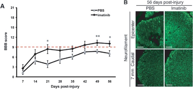
    1. Adzemovic M. Z., Zeitelhofer M., Eriksson U., Olsson T. and Nilsson I. (2013). Imatinib Ameliorates neuroinflammation in a rat model of multiple sclerosis by enhancing blood-brain barrier integrity and by modulating the peripheral immune response. PLoS ONE 8, e56586 10.1371/journal.pone.0056586 - DOI - PMC - PubMed
    1. Ahuja C. S. and Fehlings M. (2016). Concise review: bridging the gap: novel neuroregenerative and neuroprotective strategies in spinal cord injury. Stem Cells Transl. Med. 5, 914-924. 10.5966/sctm.2015-0381 - DOI - PMC - PubMed
    1. Anderson M. A., Burda J. E., Ren Y., Ao Y., O'Shea T. M., Kawaguchi R., Coppola G., Khakh B. S., Deming T. J. and Sofroniew M. V. (2016). Astrocyte scar formation aids central nervous system axon regeneration. Nature 532, 195-200. 10.1038/nature17623 - DOI - PMC - PubMed

Publication types