. 2016 Nov 21;60(12):7396-7401.
doi: 10.1128/AAC.01405-16. Print 2016 Dec.

Siderophore Cephalosporin Cefiderocol Utilizes Ferric Iron Transporter Systems for Antibacterial Activity against Pseudomonas aeruginosa

Affiliations

Siderophore Cephalosporin Cefiderocol Utilizes Ferric Iron Transporter Systems for Antibacterial Activity against Pseudomonas aeruginosa

Akinobu Ito et al. Antimicrob Agents Chemother. .

Abstract

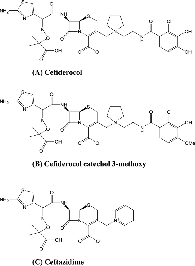
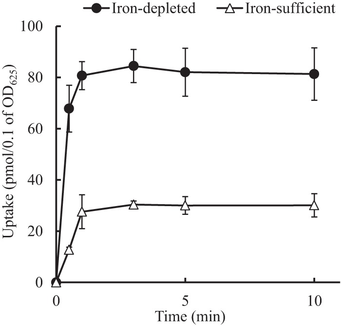
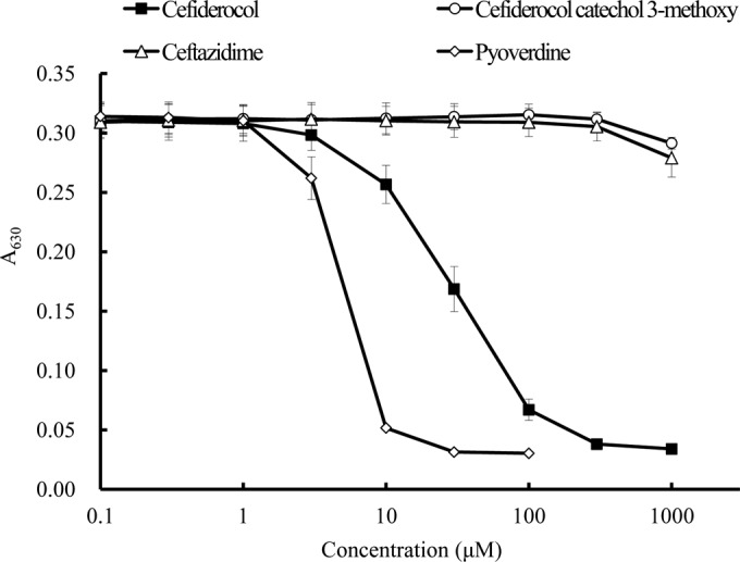
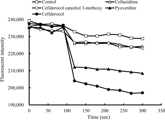
Cefiderocol (S-649266) is a novel parenteral siderophore cephalosporin conjugated with a catechol moiety at the third-position side chain. The in vitro activity of cefiderocol against Pseudomonas aeruginosa was enhanced under iron-depleted conditions, whereas that of ceftazidime was not affected. The monitoring of [thiazole-14C]cefiderocol revealed the increased intracellular accumulation of cefiderocol in P. aeruginosa cells incubated under iron-depleted conditions compared with those incubated under iron-sufficient conditions. Cefiderocol was shown to have potent chelating activity with ferric iron, and extracellular iron was efficiently transported into P. aeruginosa cells in the presence of cefiderocol as well as siderophores, while enhanced transport of extracellular ferric iron was not observed when one of the hydroxyl groups of the catechol moiety of cefiderocol was replaced with a methoxy group. We conclude that cefiderocol forms a chelating complex with iron, which is actively transported into P. aeruginosa cells via iron transporters, resulting in potent antibacterial activity of cefiderocol against P. aeruginosa.


Figures

FIG 1
FIG 1
Chemical structures of cefiderocol, cefiderocol catechol 3-methoxy, and ceftazidime.
FIG 2
FIG 2
Uptake of [thiazole-14C]cefiderocol into P. aeruginosa PAO1. The test strain was incubated under iron-depleted (ID-CAMHB) and iron-sufficient [ID-CAMHB supplemented with 100 μM ammonium iron(III) citrate] conditions overnight. After the culture was washed and incubated at 37°C for 10 min, 66 μM ammonium iron(III) citrate and 10 μg/ml [thiazole-14C]cefiderocol were simultaneously added. Samples were taken at 0.5, 1, 3, 5, and 10 min and filtered, and the radioactivity was determined by a liquid scintillation counter. The uptake amounts of [thiazole-14C]cefiderocol were significantly different between the cells grown in ID-CAMHB and those grown in CAMHB at all time points (P value < 0.05, by t test).
FIG 3
FIG 3
Chelating activity of cefiderocol with ferric iron determined by chrome azurol B. The chelating activity was determined by the color change of chrome azurol B in the presence of cetyltrimethylammonium bromide. The color change was detected by measuring the A630 with a spectrophotometer. The chelating activities were significantly different between cefiderocol and ceftazidime and between cefiderocol and cefiderocol catechol 3-methoxy at concentrations of 10, 30, 100, 300, and 1,000 μM (P value < 0.05, by t test). The chelating activities were also significantly different between pyoverdine and ceftazidime and between pyoverdine and cefiderocol catechol 3-methoxy at concentrations of 3, 10, 30, and 100 μM (P value < 0.05, by t test).
FIG 4
FIG 4
Iron uptake by P. aeruginosa PAO1 detected by calcein fluorescence intensity in the cells. Calcein-AM was added to the overnight culture in iron-depleted medium (ID-CAMHB); the culture was incubated for 120 min, and green fluorescence intensity was monitored by an EnVision Multilabel reader with 492-nm excitation and 535-nm emission. During 300 s of monitoring, 50 μg/ml of the compound was added 90 s after the initiation of the monitoring.

References

    1. Denis O, Nonhoff C, Dowzicky MJ. 2014. Antimicrobial susceptibility among Gram-positive and Gram-negative isolates collected in Europe between 2004 and 2010. J Glob Antimicrob Resist 2:155–161. doi:10.1016/j.jgar.2014.05.001. - DOI - PubMed
    1. Kanj SS, Whitelaw A, Dowzicky MJ. 2014. In vitro activity of tigecycline and comparators against Gram-positive and Gram-negative isolates collected from the Middle East and Africa between 2004 and 2011. Int J Antimicrob Agents 43:170–178. doi:10.1016/j.ijantimicag.2013.10.011. - DOI - PubMed
    1. Woodford N, Turton JF, Livermore DM. 2011. Multiresistant Gram-negative bacteria: the role of high-risk clones in the dissemination of antibiotic resistance. FEMS Microbiol Rev 35:736–755. doi:10.1111/j.1574-6976.2011.00268.x. - DOI - PubMed
    1. Bonomo RA, Szabo D. 2006. Mechanisms of multidrug resistance in Acinetobacter species and Pseudomonas aeruginosa. Clin Infect Dis 43:S49–S56. doi:10.1086/504477. - DOI - PubMed
    1. Farra A, Islam S, Strålfors A, Sörberg M, Wretlind B. 2008. Role of outer membrane protein OprD and penicillin-binding proteins in resistance of Pseudomonas aeruginosa to imipenem and meropenem. Int J Antimicrob Agents 31:427–433. doi:10.1016/j.ijantimicag.2007.12.016. - DOI - PubMed

MeSH terms