. 2016 Nov 18;84(12):3575-3583.
doi: 10.1128/IAI.00596-16. Print 2016 Dec.

Interleukin-17A (IL-17A) and IL-17F Are Critical for Antimicrobial Peptide Production and Clearance of Staphylococcus aureus Nasal Colonization

Affiliations

Interleukin-17A (IL-17A) and IL-17F Are Critical for Antimicrobial Peptide Production and Clearance of Staphylococcus aureus Nasal Colonization

Nathan K Archer et al. Infect Immun. .

Abstract

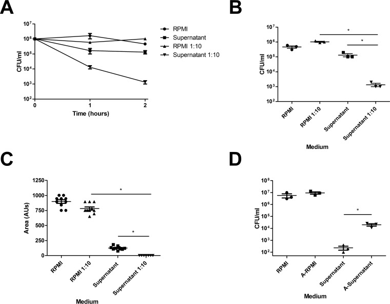
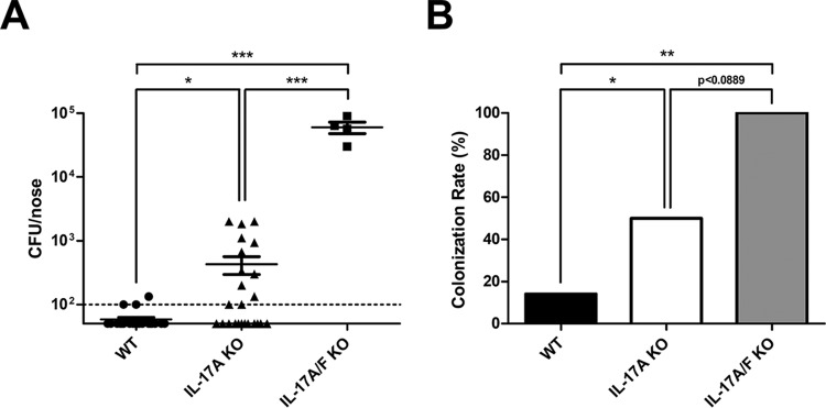
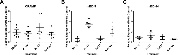
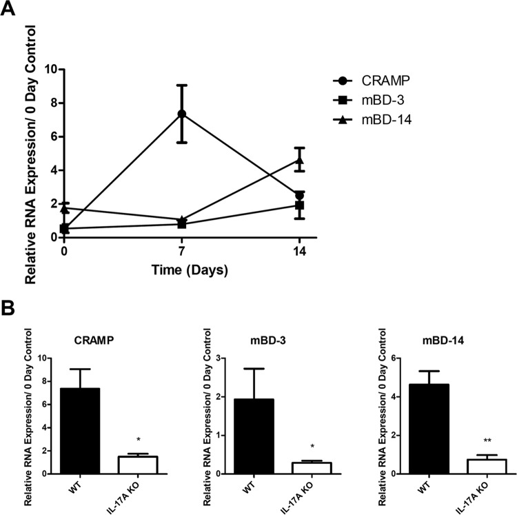
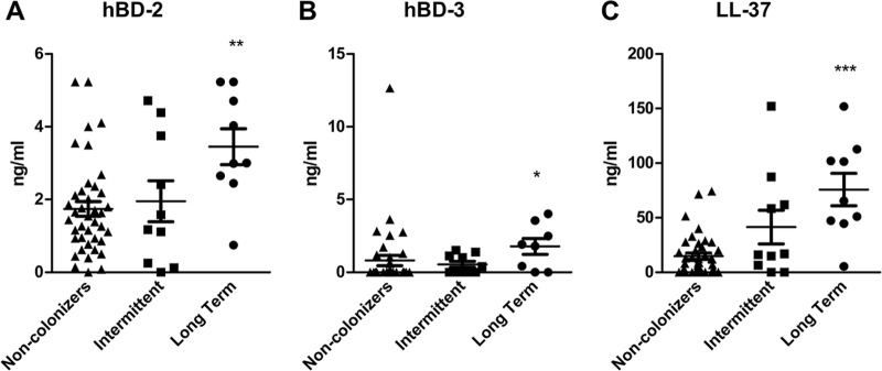
Approximately 20% of the population is persistently colonized by Staphylococcus aureus in the nares. Th17-like immune responses mediated by the interleukin-17 (IL-17) family of cytokines and neutrophils are becoming recognized as relevant host defense mechanisms for resolution of S. aureus mucocutaneous infections. Since antimicrobial peptides are regulated by the IL-17 cytokines, we sought to determine the role of IL-17 cytokines in production of antimicrobial peptides in a murine model of S. aureus nasal carriage. We discovered that nasal tissue supernatants have antistaphylococcal activity, and mice deficient in both IL-17A and IL-17F lost the ability to clear S. aureus nasal colonization. IL-17A was found to be sufficient for nasal mBD-3 production ex vivo and was required for CRAMP, mBD-3, and mBD-14 expression in response to S. aureus colonization in vivo These data were confirmed in a clinical study of nasal secretions in which elevated levels of the human forms of these antimicrobial peptides were found in nasal secretions from healthy human subjects when they were colonized with S. aureus but not in secretions from noncolonized subjects. Together, these data provide evidence for the importance of IL-17A regulation of antimicrobial peptides and IL-17F in the clearance of S. aureus nasal carriage.


Figures

FIG 1
FIG 1
Nasal tissue supernatants have antistaphylococcal activity that is solute and heat sensitive. Naive nasal tissue was harvested from WT C57BL/6J mice. Tissue was incubated in RPMI for 24 h. Tissue supernatants were utilized for in vitro killing assays. (A) S. aureus (106 CFU) was cultured in nasal tissue supernatants or supernatants diluted 1:10 in ddH2O for various amounts of time. RPMI and RPMI diluted 1:10 in ddH2O served as controls. (B) CFU counts at 2 h postinduction. (C) S. aureus was incubated for 2 h in RPMI or tissue supernatants and plated on TSA. Colony area was assessed by visualization on an Alpha Innotech FluorChem 8900 imager. AUs, arbitrary units. (D) S. aureus was incubated for 2 h in diluted RPMI or tissue supernatant or for 2 h in previously autoclaved RPMI (A-RPMI) or supernatant (A-Supernatant). n = 3 nasal tissue samples per group. Data are representative of two independent experiments. Statistical analysis was performed with a one-tailed t test (*, P < 0.05).
FIG 2
FIG 2
IL-17A and IL-17F are required for clearance of S. aureus nasal carriage. C57BL/6J WT, IL-17A KO, and IL-17A/F KO mice were inoculated intranasally with S. aureus clinical isolate SA1108. (A) CFU counts/nose 28 days postinoculation (dpi). Nasal tissue was harvested at 28 dpi, homogenized, and plated on CHROMagar-S. aureus to determine CFU counts. The detection limit was 100 CFU. (B) Colonization rate 28 days postinoculation. Colonization rate was determined by the number of mice positive for S. aureus out of the total number of mice. n = 4 to 9 C57BL/6J mice per group. Data are combined from one or two independent experiments. Statistical analyses are in comparison to results for WT mice or between groups, as indicated by lines, and were performed with a one-tailed t test or Fisher's exact test (*, P < 0.05; **, P < 0.01; ***, P < 0.001).
FIG 3
FIG 3
IL-17A is sufficient for expression of mBD-3 in ex vivo nasal tissue. Naive nasal tissue was harvested from WT C57BL/6J mice. Tissue was incubated in RPMI for 24 h with 20 ng/ml of mouse rIL-17A, rIL-17F, or both rIL-17A and rIL-17F. Nonsupplemented medium acted as a negative control. Nasal tissue RNA was purified, converted to cDNA, and used for RT-PCR assays. Levels of mouse CRAMP (A), mBD-3 (B), and mBD-14 (C) were measured. Data are presented as RNA expression relative to that of medium (nonsupplemented) controls. n = 3 to 6 C57BL/6J mice per time point. Data are representative of two independent experiments. Statistical analyses are in comparison to medium control samples and were performed with a one-tailed t test (*, P < 0.05; **, P < 0.01).
FIG 4
FIG 4
IL-17A is required for AMP expression induced by S. aureus nasal colonization. WT C57BL/6J and IL-17A KO mice were inoculated intranasally with S. aureus clinical isolate SA1108. Nasal tissue RNA was purified, converted to cDNA, and used for RT-PCR assays. (A) Levels of mouse CRAMP, mBD-3, and mBD-14 were measured in WT mice at various time points. (B) Expression levels of CRAMP, mBD-3, and mBD-14 were compared between WT and IL-17A KO mice at either day 7 or day 14. Data are presented as RNA expression relative to that of day 0 (noninoculated) controls. n = 2 to 5 C57BL/6J or IL-17A KO mice per time point. Data are representative of at least two independent experiments. Statistical analysis was performed with a one-tailed t test (*, P < 0.05; **, P < 0.01).
FIG 5
FIG 5
Human S. aureus nasal carriage promotes AMP production. Human nasal secretions were determined by culture and PCR to be either from noncolonizers, intermittent colonizers, or long-term S. aureus colonizers, and levels of hBD-2 (A), hBD-3 (B), and LL-37 (C) were elucidated by ELISAs. n = 41 noncolonizers; n = 10 intermittent colonizers; n = 9 long-term colonized human samples. Statistical analyses are in comparison to results for the noncolonizer group and were performed with the Mann-Whitney test (*, P < 0.05; **, P < 0.01; ***, P < 0.001).

References

    1. Bardan A, Nizet V, Gallo RL. 2004. Antimicrobial peptides and the skin. Expert Opin Biol Ther 4:543–549. doi:10.1517/14712598.4.4.543. - DOI - PubMed
    1. Selsted ME, Harwig SS, Ganz T, Schilling JW, Lehrer RI. 1985. Primary structures of three human neutrophil defensins. J Clin Investig 76:1436–1439. doi:10.1172/JCI112121. - DOI - PMC - PubMed
    1. Lehmann J, Retz M, Harder J, Krams M, Kellner U, Hartmann J, Hohgrawe K, Raffenberg U, Gerber M, Loch T, Weichert-Jacobsen K, Stockle M. 2002. Expression of human beta-defensins 1 and 2 in kidneys with chronic bacterial infection. BMC Infect Dis 2:20. doi:10.1186/1471-2334-2-20. - DOI - PMC - PubMed
    1. Jenssen H, Hamill P, Hancock RE. 2006. Peptide antimicrobial agents. Clin Microbiol Rev 19:491–511. doi:10.1128/CMR.00056-05. - DOI - PMC - PubMed
    1. Doss M, White MR, Tecle T, Hartshorn KL. 2010. Human defensins and LL-37 in mucosal immunity. J Leukoc Biol 87:79–92. doi:10.1189/jlb.0609382. - DOI - PMC - PubMed

LinkOut - more resources