. 2016 Oct 13;11(10):e0164588.
doi: 10.1371/journal.pone.0164588. eCollection 2016.

Characterization of the Neisseria meningitidis Helicase RecG

Affiliations

Characterization of the Neisseria meningitidis Helicase RecG

Getachew Tesfaye Beyene et al. PLoS One. .

Abstract

Neisseria meningitidis (Nm) is a Gram-negative oral commensal that opportunistically can cause septicaemia and/or meningitis. Here, we overexpressed, purified and characterized the Nm DNA repair/recombination helicase RecG (RecGNm) and examined its role during genotoxic stress. RecGNm possessed ATP-dependent DNA binding and unwinding activities in vitro on a variety of DNA model substrates including a Holliday junction (HJ). Database searching of the Nm genomes identified 49 single nucleotide polymorphisms (SNPs) in the recGNm including 37 non-synonymous SNPs (nsSNPs), and 7 of the nsSNPs were located in the codons for conserved active site residues of RecGNm. A transient reduction in transformation of DNA was observed in the Nm ΔrecG strain as compared to the wildtype. The gene encoding recGNm also contained an unusually high number of the DNA uptake sequence (DUS) that facilitate transformation in neisserial species. The differentially abundant protein profiles of the Nm wildtype and ΔrecG strains suggest that expression of RecGNm might be linked to expression of other proteins involved in DNA repair, recombination and replication, pilus biogenesis, glycan biosynthesis and ribosomal activity. This might explain the growth defect that was observed in the Nm ΔrecG null mutant.


Conflict of interest statement

The authors have declared that no competing interests exist.

Figures

Fig 1
Fig 1. Schematic diagram of model DNA substrates, and the RecGNm DNA binding and unwinding activity.
Minus and plus symbols indicate the absence or presence of RecGNm activity on the indicated substrate, respectively.
Fig 2
Fig 2. RecGNm binds different model DNA substrates.
A. Representative gel images of DNA binding assays where increasing amounts of RecGNm was incubated with i. Holliday junction, ii. lagging strand replication fork, iii. leading strand replication fork, and iv). full replication fork. Lanes 1) reaction without protein; 2–6) 25, 50, 100, 200 and 400nM RecGNm, respectively, 7) 400nM RecGNmK294A. B. Quantitation of the gel images i, ii, iii, and iv in (A.) and the calculated Kd value for each Kd = 21.42, Kd = 97.79, Kd = 216.6, and Kd = 92.18, respectively. Data presented is mean ± SD from 3 independent experiments.
Fig 3
Fig 3. RecGNm branch-migrates Holliday junction and catalyses unwinding of replication forks.
Gel images of DNA unwinding assays where increasing amounts of RecGNm was incubated with 0.1 nM A. Holliday junction, B. full replication fork, C. leading strand replication fork, D. lagging strand replication fork. Lanes: 1) reaction without enzyme, for Holliday junction substrate, 2–7) 12.5, 25, 50, 100, 200, and 400 nM RecGNm, respectively, 8) 400 nM RecGNmK294A; for fork substrates, 2–7) 1, 2, 4, 6, 12, and 25 nM RecGNm, respectively, 8) 25 nM RecGNmK294A, 9) Boiled substrate.
Fig 4
Fig 4. RecGNm is a DNA dependent ATPase.
A graph showing ATPase activity of RecG from Neisseria meningitidis (RecGNm) and RecGNmK294A in the presence of DNA cofactors; forked DNA duplex, leading strand replication fork, lagging strand replication fork, HJ, M13mp18 ssDNA, and pET28b(+) dsDNA. % ATP hydrolysis is a measure of the percentage of inorganic phosphate released by the cleavage of the γ-phosphate of ATP. The standard deviations indicated by bars are from 3 independent experiments.
Fig 5
Fig 5. Phenotypic characterization of the NmΔrecG mutant.
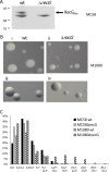
A. Immunoblot of cell lysates from Neisseria meningitidis (Nm) Mc58 wildtype and a Mc58ΔrecG mutant detected with anti-RecG antiserum. B. Colony morphology of M1080 wildtype and M1080ΔrecG null mutant of grown overnight at 34°C, showing normal size colonies for the wildtype (i and iii), whereas the mutant strain shows normal size colonies together with small size colonies (ii and iv). C. Graphical presentation of the colony size measurement (area in mm2) of the Nm wildtype strains and NmΔrecG Mc58 and M1080 mutant strains.
Fig 6
Fig 6. DNA transformation is reduced in ΔrecG mutant.
Quantitative transformation of Neisseria meningitides with DUS-containing plasmid DNA. The standard deviations of the median from 3 independent experiments are indicated by bars. The values on the Y- axis are in logarithmic scale. Five agar plates were inoculated from each sample.
Fig 7
Fig 7. Distribution of Neisseria gonorrhoeae MS11 wildtype and ΔrecG mutant cells in flow cytometry analysis.
Flow cytometry of Hoechst-stained, fixed bacterial cells was performed. The x-axis shows fluorescence levels, which indicates the amount of DNA content per particle counted. Genome equivalents were determined from the stationary phase and rif-treated cells. The graphs represent the distribution of MS11 wildtype and ΔrecG mutant strains, A. from the exponential culture, B. treated with rifampicin and cephalexin acquired by selecting/gating the sub-population of cells/particles with fluorescence level corresponding to chromosome equivalents of 2, 4, 6, 8, 10 and 12.
Fig 8
Fig 8. Alkylating agent, DNA cross linker and UV light affect the survival of a Neisseria meningitidis ΔrecG mutant.
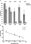
A. N. meningitidis (Nm) MC58 wildtype and MC58ΔrecG mutant were treated with hydrogen peroxide, paraquat, MMS and MMC. B. Survival rate of Nm MC58 wildtype and MC58ΔrecG was determined after exposing the cells to the indicated UV influences. The standard deviations of the median from 3 independent experiments are indicated by bars.
Fig 9
Fig 9. Neisseria meningitidis recG (recGNm) is a DNA uptake sequence (DUS) abundant gene.
A. Schematic diagram of the N. meningitidis (Nm) recG gene (recGNm) and neighbouring genes showing the position of the DNA uptake sequences (DUS) (black arrows). B. Domain organization in the RecGNm and non-synonymous single nucleotide polymorphism (nsSNP) identified in recGNm from different Nm strains. The positioning of the nsSNPs is shown in square tick marks (bold). C. The predicted structure of RecGNm with colour coding for conserved (red) and variable (blue) regions, yellow regions indicate insufficient data. The RecGNm regions outside of the helicase motifs are shown in transparent.
Fig 10
Fig 10. Functional classification of proteins identified and differentially expressed in Neisseria meningitidis by mass spectrometry.
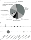
A. Pie-chart representing the functional classification of all identified proteins identified using high-resolution mass spectrometry. The 1073 proteins were distributed in 10 functional categories based on KEGG orthology using BlastKOALA. B. Differentially more abundant and less abundant proteins in Nm wildtype and NmΔrecG sorted by KEGG where the dot plot size is proportional to the counts of differentially expressed (DE) proteins.
Fig 11
Fig 11. RecGNm directly interacts with SSBNm.
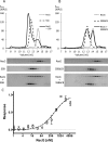
A. Co-gel filtration analysis was performed to monitor the interaction between of RecGNm and SSBNm. Upper panels: chromatogram A280 [mAU] vs retention volume [ml]. Lower panels: SDS-PAGE of 13μl sample from each 0.5ml fraction beginning from 9.5 ml up to 16 ml, and stained with Coomassie blue. A. 20 μM RecGNm mixed with 40 μM SSBNm and each protein alone. B. 20 μM RecGNm mixed with 40 μM SSB NmΔC8 and each protein alone. C. Microscale thermophoresis (MST) analysis of the interaction between RecGNm and SSBNm. MST results of three independent experiments were included. The average and standard deviation of the normalised response and the fitted curve are shown. The calculated Kd value of the interaction is 558 ±139 nM.

References

    1. Nassif X (2000) Microbiology. A Furtive Pathogen Revealed. Science (80-) 287: 1767–1768. doi: 10.1126/science.287.5459.1767 - DOI - PubMed
    1. Davidsen T, Tønjum T (2006) Meningococcal genome dynamics. Nat Rev Microbiol 4: 11–22. doi: 10.1038/nrmicro1324 - DOI - PubMed
    1. Black CG, Fyfe JA, Davies JK, Black CG, Janet AM, Fyfe JKD (1998) Absence of an SOS-like system in Neisseria gonorrhoeae. Gene 208: 61–66. - PubMed
    1. Nagorska K, Silhan J, Li Y, Pelicic V, Freemont PS, Baldwin GS, et al. (2012) A network of enzymes involved in repair of oxidative DNA damage in Neisseria meningitidis. Mol Microbiol 83: 1064–1079. doi: 10.1111/j.1365-2958.2012.07989.x - DOI - PMC - PubMed
    1. Brosh RM (2013) DNA helicases involved in DNA repair and their roles in cancer. Nat Rev Cancer 13: 542–558. doi: 10.1038/nrc3560 - DOI - PMC - PubMed

MeSH terms

LinkOut - more resources