Review
. 2016 Aug;1378(1):158-165.
doi: 10.1111/nyas.13226.

The molecules in the corneal basement membrane zone affected by mustard exposure suggest potential therapies

Affiliations
Review

The molecules in the corneal basement membrane zone affected by mustard exposure suggest potential therapies

Marion K Gordon et al. Ann N Y Acad Sci. 2016 Aug.

Abstract

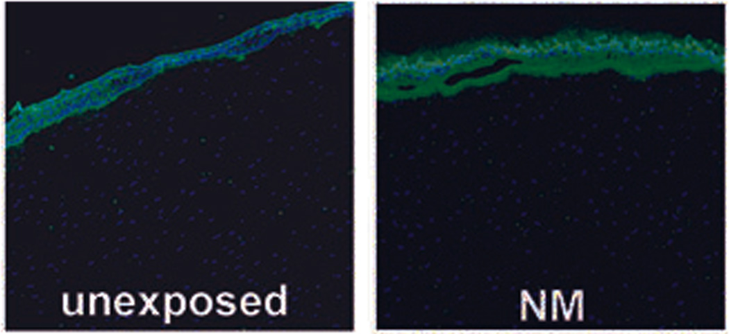
Mustard exposures result in epithelial-stromal separations in the cornea and epidermal-dermal separations in the skin. Large blisters often manifest in skin, while the cornea develops microblisters, and, when enough form, the epithelium sloughs. If the exposure is severe, healing can be imperfect and can result in long-term adverse consequences. For the cornea, this could manifest as recurrent corneal erosions. Since the corneal epithelial-stromal separations are in the region identified by electron microscopy as the lamina lucida, the same region affected by the blistering disease junctional epidermolysis bullosa (JEB), we postulated that the molecules that are defective in JEB would be the same ones cleaved by mustard compounds. These molecules are α6β4 integrin and collagen XVII, which can be cleaved by matrix metalloproteinase-9 (MMP-9) and ADAM17, respectively. Therefore, our laboratory has tested MMP-9 and ADAM17 inhibitors as potential therapies to attenuate corneal mustard injury. Our results demonstrated that inhibiting MMP-9 and ADAM17 resulted in less epithelial-stromal separation in the corneas at 24 h postexposure, as compared with using only medium as a therapy.

Keywords: basement membrane zone; collagen XVII; cornea; mustard injury; mustard therapies; α6β4 integrin.


Conflict of interest statement

The authors declare no conflicts of interest.

Figures

Figure 1
Figure 1
Comparison of unexposed corneas with those with injury caused by NM and SM. Both vesicants induce separation at the epithelial–stromal border, starting with microblisters (D and G). When these coalesce, they progress to larger areas of epithelial–stromal separation (E and H). Eventually the epithelium loses the last tendrils holding on to the stroma, and the epithelial cell layer is released (F and I). Reprinted from Ref. .
Figure 2
Figure 2
Electron micrographs of the epithelial–stromal border in an unexposed organ-cultured rabbit eye (A) and a freshly dissected rabbit eye (B). Note that the hemidesmosomes are flattened at the basal epithelial cell surface (A and B). In corneal organ cultures exposed to NM (C and D), and in rabbit eyes exposed in vivo to SM (E and F), the hemidesmosomes are not flat and sometimes appear quite plump. The lamina luicida after exposure to either vesicant is expanded between the hemidesmosomes, and in some micrographs thin tendrils can be seen reaching from the hemidesmosomes toward the stromal surface (C, D, E, and F). Reprinted from Ref. .
Figure 3
Figure 3
MMP-9 immunofluorescence (IF): unexposed corneal organ cultures have very low levels of MMP-9 expression as assessed by IF (left panel). At 22 h after a 2-h exposure to NM, MMP-9 is very abundant at the epithelial–stromal border. Reprinted from Ref. .
Figure 4
Figure 4
An unexposed rabbit eye (top); the central three vertical images are in vivo SM-exposed rabbit eyes at 1 day, 3 days, and 7 days postexposure that received no treatment, and the bottom three horizontal images are SM-exposed eyes at 28 days postexposure. Note that the SM-exposed cornea receiving no therapy (left) is still cloudy, while the middle and right photos are eyes treated with doxycycline and are fairly well healed. There is some evidence of neovascularization, but less in the SM-exposed eye receiving no therapy. Reprinted from Ref. .
Figure 5
Figure 5
(A) A mouse monoclonal antibody against ADAM17 detected no rabbit ADAM17 in unexposed corneas by immunofluorescence (left panel), but did detect it in NM-exposed corneas (right panel). Both had been cultured for 24 h. (B) The Innozyme TACE activity assay kit showed unexposed corneas kept in culture for 24 h show very little ADAM17 activity (~40 ng/mL) (left panel), while corneas exposed to NM for 2 h then kept in culture for 22 h showed ~ 24 times more ADAM17 activity than the unexposed corneas kept in culture for the same amount of time (right panel). (C) The Innozyme TACE activity kit determined how fast ADAM17 was turned on by NM. When applied and immediately washed off (0 min), or allowed to remain on the cornea for 5 or 10 min, ADAM17 assays indicated the enzyme was immediately activated. Thus, ADAM17 activation is an early response to NM exposure (left panel) Epithelial–stromal separation averaged 85% in the sets of corneas receiving no therapy, but with three applications of the hydroxamate compound NDH4417, separation was only ~17%. (right panel). The statistical analysis included one-way analysis of variance followed by Duncan's multiple comparison tests, then Duncan’s multiple comparison test or the unpaired Student’s t-test. All data published in Ref. . *A P value < 0.05 was considered significant.

References

    1. Graham JS, Schoneboom BA. Historical perspective on effects and treatment of sulfur mustard injuries. Chemico-Biological Interactions. 2013;206:512–522. - PubMed
    1. Gordon MK, Enzenauer RW, Babin MC. Ocular Toxicity of Sulfur Mustard. In: Gupta RC, editor. Handbook of Toxicology of Chemical Warfare Agents. Chapter 39. 2009. pp. 575–594.
    1. The Guardian. UN watchdog confirms mustard gas attack in Syria Organisation for the Prohibition of Chemical Weapons says child was probably filled by mustard gas in August during fighting between jihadi and rebels. [Accessed April 10, 2016];2015 Nov 6; http://www.theguardian.com/world/2015/nov/06/un-watchdog-confirms-mustar....
    1. Boston Globe. Reported by Helene Cooper and Eric Schmitt. ISIS chemical weapons specialist details militants’ plans, US says. [Accessed August 16, 2016];2016 Mar 9; https://www.bostonglobe.com/news/world/2016/03/09/captures-head-isis-che....
    1. Solberg Y, Alcalay M, Belkin M. Ocular injury by mustard gas. Surv. Ophthalmol. 1997;41:461–466. - PubMed

Publication types

MeSH terms

Substances

LinkOut - more resources